Computed tomography (CT) imaging is a medical diagnostic technique that uses X-rays and computer processing to create detailed cross-sectional images of the body. Unlike conventional radiography, which produces a single two-dimensional projection, CT captures multiple X-ray measurements from different angles around the patient. These data are then reconstructed by powerful algorithms into slices that can be viewed individually or combined to form three-dimensional representations of anatomical structures.
CT imaging is widely used in clinical practice because it can reveal internal details with high clarity, enabling accurate assessment of organs, blood vessels, bones, and soft tissues. It is a valuable tool in the detection and monitoring of conditions such as tumours, vascular diseases, trauma, and infections. By offering rapid image acquisition and the ability to guide interventions, CT has become integral to emergency medicine, oncology, cardiology, and many other specialties.
CT Scenario Video and Knowledge Check
Watch the computed tomography (CT) video before attempting the scenario quiz. In this training scenario, a patient is referred to a large teaching hospital for CT imaging. The video walks you through the workflow and explains key concepts—attenuation, reconstruction, windowing, pitch, dose optimisation, and contrast administration—showing how they are applied in clinical practice. At the end, complete the knowledge-check quiz. The questions are drawn directly from the video, so pay close attention to the details as you progress.
Scenario: Sarah’s CT Examination
Referral for imaging
Sarah, a 52-year-old woman, presented at the emergency department with acute abdominal pain. After initial assessment, the clinician requested an abdominal CT scan to rule out causes such as appendicitis, perforation, or bowel obstruction. This is a common indication for CT imaging, since the technique can rapidly visualise internal organs, vascular structures, and free air.
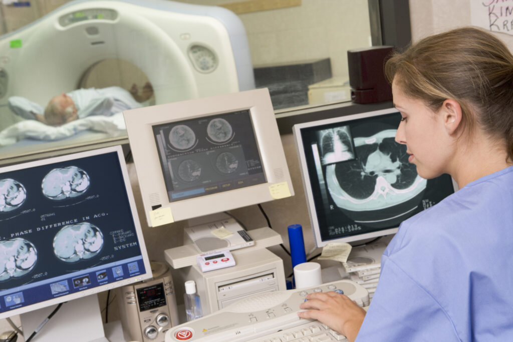
The CT scanner environment
Sarah was taken to the radiology department. The radiographer explained that she would be positioned on the patient table, which would pass through the CT gantry. The gantry houses the X-ray tube, detectors, and the rotating mechanical system required to generate the images.
As the table moved into position, the radiographer reassured her that the scan would be painless but involved exposure to ionising radiation—a typical risk of CT.
Axial imaging and display
The scan was performed in axial mode, where images are acquired as cross-sections perpendicular to the patient’s long axis. These axial slices were then reconstructed and displayed on the workstation. Radiologists typically review CT images in axial format, although multiplanar reformats (MPRs) and 3D renderings are also possible.
Tissue Attenuation and Hounsfield Units
On the workstation, the radiographer reviewed images measured in Hounsfield Units (HU)—the scale used to quantify tissue attenuation in CT. Water has a HU value of 0, and all other tissues are measured relative to this. For example:
- Lung tissue typically measures around -800 to -900 HU.
- Fat measures around -100 HU.
- Soft tissues such as liver or muscle measure around +40 HU.
- Bone can measure above +1000 HU, making it appear brightest on the scan.
This standardised scale allows radiologists to distinguish between different tissues.
Windowing in CT
The radiologist adjusted the window width and window level on the images. Windowing controls the contrast and brightness applied to CT images, enabling optimal visualisation of different tissues. A narrow window is used for soft tissues, while a wide window is applied for lung or bone to prevent saturation of the bright or dark areas.
Use of contrast media
For Sarah’s case, the radiologist required intravenous iodinated contrast media, the most common type used in CT. Contrast improves visualisation of vascular structures and enhances differentiation between normal and abnormal tissue. For example, contrast can highlight a perforated bowel or detect abnormal vascular patterns. Without contrast, some pathologies would be difficult to identify.
Multislice CT
The scanner used was a 64-slice multislice CT system. Multislice CT refers to scanners with multiple rows of detectors, allowing acquisition of many slices in a single rotation. This enables faster scanning, improved spatial resolution, and reduced motion artefacts, which is particularly useful in abdominal and cardiac imaging.
Helical scanning and pitch
During Sarah’s examination, the table moved continuously as the gantry rotated, performing a helical CT scan. In this mode, the concept of pitch becomes important. Pitch is the ratio of table movement per gantry rotation to the beam width. A higher pitch means faster scanning and potentially lower dose, but with some loss of image resolution. A lower pitch provides higher resolution but increases dose and scan time.
Image reconstruction
The acquired raw data were processed using iterative reconstruction algorithms. Iterative reconstruction reduces image noise and improves quality compared with traditional filtered back projection, especially at lower radiation doses. This allows the department to achieve diagnostic-quality images while keeping patient dose as low as reasonably achievable (ALARA).
Beam hardening artefacts
As the radiologist scrolled through the images, they noted mild streak artefacts adjacent to dense bony structures. This was explained by beam hardening—an artefact caused when lower-energy X-ray photons are absorbed more readily as the beam passes through dense material such as bone, leaving the beam “harder” (with higher average energy). Corrections in software help reduce this effect, but it remains a familiar artefact in CT imaging.
Dose considerations and radiosensitive organs
Although CT is invaluable for diagnosis, it involves higher doses than conventional radiography. In abdominal CT, the radiosensitive organs of concern include the ovaries in women and the testes in men. In general, the most radiosensitive organ in CT is the thyroid gland, though breast and gonadal tissues are also highly sensitive. Dose optimisation techniques such as automatic tube current modulation, shielding, and iterative reconstruction all contribute to reducing unnecessary exposure.
Final diagnosis
The radiologist completed the review of Sarah’s images. The contrast-enhanced CT clearly demonstrated an inflamed appendix with peri-appendiceal fat stranding, consistent with acute appendicitis. Thanks to rapid imaging, Sarah was able to proceed quickly to surgery with confidence in the diagnosis.
Key Concepts Illustrated
Through Sarah’s CT examination, several fundamental concepts were demonstrated:
- CT stands for Computed Tomography, a cross-sectional imaging technique using X-rays.
- Images are measured in Hounsfield Units, with water at 0 HU.
- Axial slices are the standard format of CT display.
- Windowing controls image contrast and brightness.
- Iodinated contrast media enhance vascular and tissue visibility.
- Bone appears brightest due to high attenuation.
- Multislice CT enables faster acquisition and improved resolution.
- Helical pitch influences scan speed, resolution, and dose.
- Iterative reconstruction improves image quality while lowering dose.
- Beam hardening is a common artefact caused by dense structures.
- The thyroid is among the most radiosensitive organs in CT.
- Radiation exposure remains a risk, so dose optimisation is essential.
Conclusion
This clinical scenario demonstrates how CT combines technology, physics, and clinical application to provide rapid, detailed imaging of the body. By understanding principles such as attenuation, windowing, contrast use, pitch, and reconstruction, radiographers and radiologists can optimise image quality while minimising patient risk.
Knowledge Check
You have now reviewed the essential principles of CT imaging through a practical clinical case. The following knowledge check quiz is designed to test your understanding of the key topics introduced in this scenario, including image reconstruction, beam hardening, windowing, contrast media, attenuation values, and dose considerations.
Instruction: Select the best answer from the options provided. Refer back to the scenario if needed, and use it to guide your responses. Completing the quiz will help you consolidate your knowledge and ensure you are confident in applying these CT concepts to clinical practice.
Disclaimer
This clinical scenario is provided for educational and training purposes only. It is a fictional case study designed to illustrate the principles and practical applications of Computed Tomography (CT) imaging in a clinical setting. The information should not be interpreted as medical advice or used as a substitute for professional diagnosis, treatment, or patient care. Healthcare professionals should always rely on their clinical judgement, institutional protocols, and current guidelines when making decisions about imaging or treatment. Any resemblance to actual persons, living or deceased, is purely coincidental.