


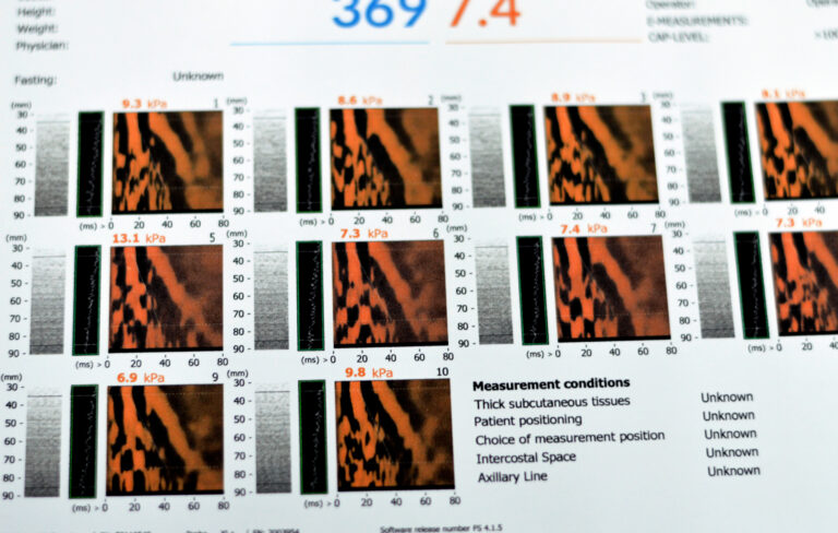
Ultrasound Elastography: Non-Invasive Evaluation of Liver Fibrosis in Chronic Disease
Understand the advantages of ultrasound elastography liver fibrosis over traditional biopsy in assessing chronic liver disease with precision.

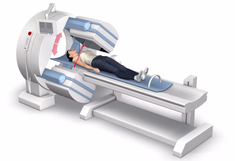
The Role of Magnets in Modern Medicine: Applications and Innovations
Magnetics in medicine enhance diagnostic precision, therapeutic targeting, and innovative treatments, transforming modern healthcare practices globally.
Gamma Radiation: Its Discovery, Applications, and Safety Considerations
Gamma radiation is essential for medical imaging, cancer treatment, industrial testing, yet requires stringent safety measures due to risks

Ensuring Patient Data Security in Remote Patient Monitoring
Remote patient monitoring transcends traditional medical settings, leveraging technology to secure data and enhance healthcare delivery.
Non-Veg Items Are Effective For Men’s Health
Discover the importance of non-veg items for men’s health, including heart-healthy fish and nutrient-rich meats.

Top Breakthroughs in Cancer Diagnosis and Prevention
Breakthroughs in cancer diagnosis and prevention now enable earlier detection, personalised care, and improved patient outcomes worldwide.