MRI is one of the most powerful tools in diagnostic imaging. Its ability to produce high-resolution images without ionising radiation has transformed the management of neurological, musculoskeletal, and oncological conditions. However, the technology behind MRI is complex. Parameters such as relaxation times, field strength, gradient performance, and sequence choice all influence image quality and diagnostic yield.
This scenario follows a patient’s journey through MRI, guided by an experienced radiologist and MRI technologist. As the case unfolds, various aspects of MRI technology, physics, safety, and artefacts are discussed in detail, providing the foundation for a later knowledge check quiz.
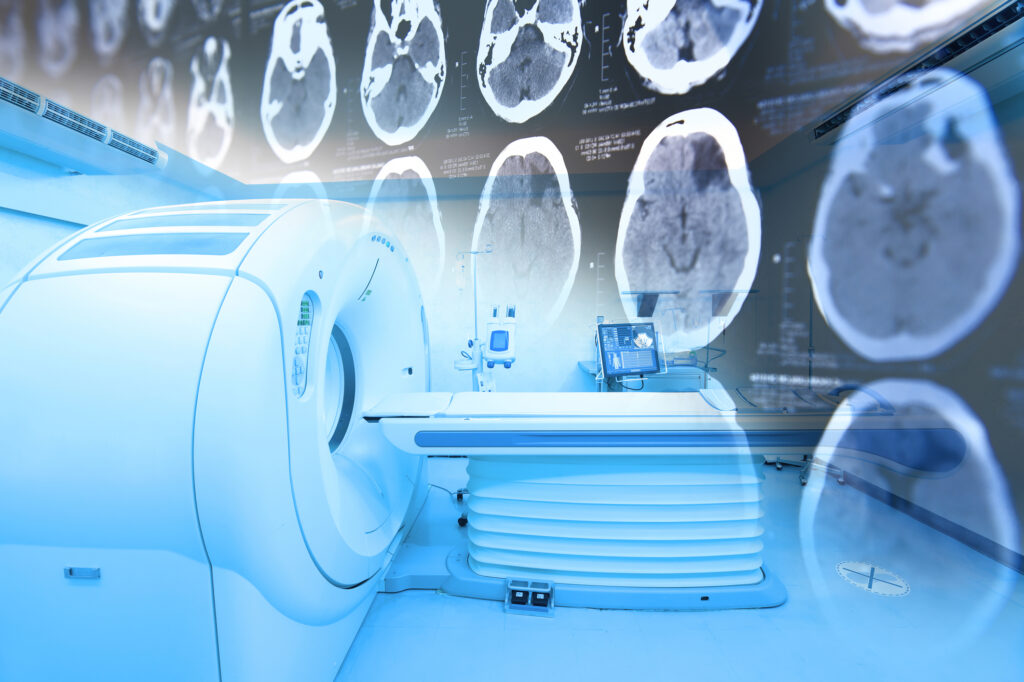
Scenario: A Teaching Session in the MRI Suite
The Patient
Mr. Collins, a 52-year-old teacher, had been referred to the hospital with intermittent headaches, mild memory lapses, and dizziness. His physician suspected a neurological cause and requested an MRI scan of the brain. He was otherwise fit and healthy, with no previous surgical history, which made him a straightforward candidate for MRI.
On the day of the scan, Dr. Wilson, a consultant neuroradiologist, invited a group of radiology trainees to join the session. Mr. Collins’s case would serve not only as a diagnostic procedure but also as a teaching opportunity.
Entering the MRI Environment
The group entered the controlled MRI suite. Dr. Wilson explained that the scanner in use was a 3 Tesla system, the standard for advanced neuroimaging in most large hospitals. She contrasted this with 7 Tesla ultra-high-field scanners, which were being introduced in research centres. These higher Tesla machines provide improved spatial resolution and signal-to-noise ratio, enabling detailed visualisation of fine brain structures. However, they also present challenges such as increased artefacts and stricter safety considerations.
MRI Safety Considerations
Before Mr. Collins entered the scanner, the MRI technologist conducted a safety screening. Dr. Wilson explained the importance of this step. Any metallic implants, pacemakers, or certain aneurysm clips could pose serious risks inside the magnet. Ferromagnetic objects can be displaced or heated by the strong magnetic field, and even non-ferromagnetic devices may malfunction.
The group noted that Mr. Collins had no implants or contraindications, making him suitable for the scan. Dr. Wilson emphasised that MRI is a non-ionising modality, unlike CT or X-ray. This is one reason why it is particularly suitable for repeated imaging and research in sensitive populations.
Physics Principles
As the technologist positioned Mr. Collins on the patient table, Dr. Wilson revisited the fundamentals. MRI relies on the principle of nuclear magnetic resonance, in which atomic nuclei align with a strong magnetic field and can be manipulated using radiofrequency pulses.
The hydrogen nucleus (proton) is the most commonly used in clinical imaging because it is abundant in the body and has a magnetic moment suitable for detection. When a radiofrequency (RF) pulse is applied, the protons are excited; as they relax back to equilibrium, they emit signals that are detected and converted into images.
The group also reviewed the role of the RF coil, which transmits the excitation pulses and detects the returning signal. Different coils — head, spine, cardiac — are designed to optimise signal collection for specific anatomical regions.
Relaxation Times and Imaging Parameters
Dr. Wilson explained that relaxation processes largely determine MRI contrast. T1 relaxation time refers to the time it takes for protons to realign with the main magnetic field, while T2 relaxation time reflects the loss of coherence among spinning protons in the transverse plane.
Parameters such as TR (repetition time) and TE (echo time) control how the scanner samples these relaxation properties. TR is the time between successive RF pulses, and TE is the time between the initial RF pulse and the signal echo detection. By manipulating TR and TE, radiologists can produce images with T1-weighted, T2-weighted, or proton-density contrast.
MRI Sequences in Practice
For Mr. Collins’s brain scan, multiple sequences were planned. T1-weighted sequences were used for anatomical detail, while T2-weighted sequences highlighted fluid and pathological changes.
Dr. Wilson highlighted the use of FLAIR (Fluid-Attenuated Inversion Recovery), which suppresses cerebrospinal fluid (CSF) signals, allowing lesions near the ventricles to be more visible. This sequence is particularly valuable in conditions such as multiple sclerosis.
Another sequence discussed was Gradient Echo (GRE), which is highly sensitive to magnetic susceptibility effects. This makes it particularly effective for detecting haemorrhage or calcification.
Diffusion-weighted imaging (DWI) was also performed. This sequence is sensitive to the random motion of water molecules in tissues and is crucial for detecting acute ischaemic stroke, as areas of restricted diffusion appear hyperintense.
Finally, Dr. Wilson explained functional MRI (fMRI), which is often used in research and pre-surgical planning. fMRI detects brain activity by measuring changes in blood oxygenation (BOLD signal). Although not needed for Mr. Collins, it was introduced as an important tool in mapping brain function.
Artefacts in MRI
As images appeared on the console, the trainees noticed small distortions. Dr. Wilson explained that MRI is prone to various artefacts. Chemical shift artefact occurs because fat and water protons resonate at slightly different frequencies, creating misregistration at fat-water interfaces. Motion artefacts, on the other hand, are common when patients move during acquisition, causing blurring or ghosting across the image.
Recognising these artefacts is essential for accurate interpretation and for deciding when a sequence should be repeated.
Gadolinium Contrast Agents
After initial sequences, Mr. Collins was administered a gadolinium-based contrast agent to assess for abnormal enhancement. Dr. Wilson explained that gadolinium primarily shortens T1 relaxation time, causing tissues that take up the agent (such as tumours or inflamed regions) to appear bright on T1-weighted images.
Gadolinium is generally safe but should be used cautiously in patients with severe renal impairment due to the risk of nephrogenic systemic fibrosis.
Gradient Coils and Image Formation
The group then discussed how spatial encoding in MRI is achieved. Gradient coils vary the magnetic field across the patient, allowing signals to be localised in three dimensions. Without gradients, MRI would not be able to produce images but only bulk signal measurements.
Field Strength and Clinical Context
While Mr. Collins’s scan was performed on a 3 Tesla scanner, Dr. Wilson reminded the trainees that many clinical scanners worldwide still operate at 1.5 Tesla. These systems remain the clinical workhorse, providing excellent diagnostic capability with fewer artefacts than higher-field systems.
Ultra-high-field systems, such as 7 Tesla MRI, are still largely confined to research environments, but they offer exciting potential for ultra-detailed neuroimaging.
Results and Interpretation
Mr. Collins’s images revealed enlarged lymph nodes at the base of the skull but no acute haemorrhage, stroke, or mass lesion. The contrast-enhanced images provided further clarity, and the diffusion sequences excluded recent ischaemia.
The radiology trainees discussed how each sequence had contributed: T1-weighted scans gave excellent structural detail; T2 and FLAIR highlighted areas of oedema; gradient echo ruled out haemorrhage; diffusion ruled out stroke; and contrast-enhanced T1 clarified vascularised pathology.
Session Conclusion
By the end of the teaching session, the trainees had not only followed a patient’s imaging journey but also revisited the foundations of MRI. They had explored:
- The principles of nuclear magnetic resonance
- The use of hydrogen protons in clinical imaging
- Relaxation times and the role of TR and TE
- Common sequences such as T1, T2, FLAIR, GRE, DWI, and fMRI
- Artefacts, including chemical shift and motion
- The function of RF and gradient coils
- The clinical applications of gadolinium contrast
- Safety concerns with metallic implants and the non-ionising nature of MRI
- The differences between 1.5T, 3T, and 7T systems
The case of Mr. Collins illustrated how MRI combines physics, technology, and clinical reasoning to provide unparalleled insight into brain pathology.
Transition to the Quiz
To consolidate their learning, the trainees were invited to complete an MRI Knowledge Check Quiz based on this scenario. The quiz would test their understanding of MRI physics, safety, sequences, artefacts, and clinical applications.
This exercise was not designed as a formal examination but as a reflective self-assessment. By working through the quiz, the trainees could identify areas for revision, confirm their grasp of fundamental principles, and strengthen their ability to apply MRI knowledge to clinical practice.
Knowledge Check
Disclaimer
The content presented in Inside the Magnet: An MRI Case Study is intended solely for educational and training purposes. The patient case, characters, and scenario are fictional and do not represent real individuals. Any resemblance to actual persons, living or deceased, is coincidental.
This material should not be used as a substitute for professional medical advice, diagnosis, or treatment. While the case study discusses technical aspects of MRI physics, safety, and clinical applications, it is not exhaustive and may omit certain considerations relevant to clinical practice. Healthcare professionals should always rely on their own training, institutional guidelines, and professional judgement when interpreting MRI scans or making clinical decisions.
The authors and publishers accept no liability for any loss, damage, or injury arising from the use of this material in clinical, educational, or research settings.
home »