X-rays are among the earliest and most widely adopted imaging techniques in medical practice. First discovered in 1895 by the German physicist Wilhelm Conrad Röntgen, their ability to create images of internal body structures without the need for surgery transformed medicine almost overnight. This breakthrough allowed clinicians to detect fractures, locate foreign objects, and diagnose a wide range of diseases with unprecedented accuracy. Over the decades, technological advances have refined X-ray equipment, reducing radiation doses and improving image quality. Today, X-rays remain a first-line diagnostic tool for many conditions, particularly in chest imaging for lung and heart assessment, skeletal imaging for fractures and joint evaluation, and abdominal imaging to detect obstructions or calcifications. Their speed, relatively low cost, and broad availability make them indispensable in both emergency and routine clinical settings, where timely diagnosis can be critical to patient outcomes.
Chest X-ray Case Study: Video and Quiz
Before starting, watch the video in full and review the clinical scenario. It follows Sarah, a 62-year-old woman admitted with chest pain and shortness of breath. Through her X-ray journey, you will see how physics, anatomy, and clinical interpretation work together in real practice. Once you have completed the video and scenario, you will be ready to answer the quiz, which covers the key concepts, findings, and techniques demonstrated.
Scenario: Sarah’s Chest and Skeletal X-rays
Clinical Presentation
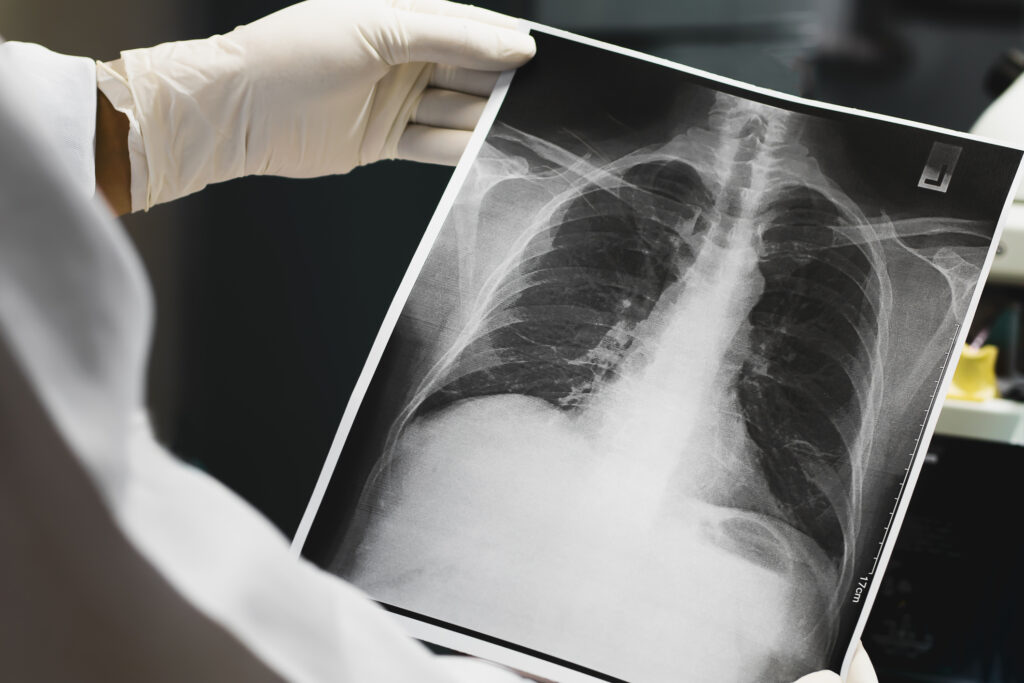
Sarah arrived in the emergency department with acute chest pain and mild breathlessness. The physician requested an urgent chest X-ray to rule out pneumonia, pleural effusion, or cardiac enlargement.
Physics and Appearance of Structures
The radiographer explained to a trainee observing the case that X-ray imaging is based on the differential attenuation of X-ray beams by various tissues:
- Dense structures like bone absorb more X-rays and appear white (radiopaque).
- Soft tissues and organs show up in varying shades of grey.
- Air-filled structures (lungs, bowel) absorb the least and appear black (radiolucent).
This principle helps distinguish anatomical structures and detect abnormalities.
Chest X-ray Technique
Sarah’s chest X-ray was performed in a posterior-anterior (PA) view, the most common projection for evaluating the lungs, heart, and mediastinum.
The radiographer reminded the trainee that:
- Adequate inspiration is confirmed when at least 10 posterior ribs are visible.
- Proper centring is assessed by checking the clavicles and trachea.
- Comparing current films with previous X-rays is vital to detect subtle changes and monitor disease progression.
Interpretation of Findings
The radiologist reviewed Sarah’s X-ray with the following observations:
- Blunting of the costophrenic angle on the right side, suggestive of a small pleural effusion.
- The silhouette sign was absent, meaning cardiac borders remained visible, helping to exclude consolidation in the adjacent lung.
- No evidence of ground-glass opacity, which would suggest interstitial or early alveolar disease.
- Heart size appeared normal, remaining within the upper limit of 50% of thoracic width.
Skeletal X-ray
Later, Sarah also reported wrist pain after a recent fall. A skeletal X-ray of the wrist was performed.
The radiographer discussed with the trainee:
- The term “fracture line” refers to the disruption of bone continuity.
- A scaphoid fracture can sometimes be difficult to detect, so special views and follow-up imaging may be required.
- Comparing both wrists can aid in identifying subtle injuries.
Dense bone appeared white, while the surrounding soft tissues were grey. A small fracture was suspected but required a second opinion from the consultant radiologist.
Artefacts and Contrast
The trainee asked about artefacts. The radiographer explained:
- Motion artefact can mimic pathology if the patient moves during exposure.
- External objects like clothing, ECG leads, or jewellery can obscure findings.
The radiologist reinforced that artefacts must always be ruled out before confirming pathology.
Contrast in X-ray imaging is influenced primarily by kilovoltage (kVp). Lower kVp increases contrast but also raises patient dose, while higher kVp reduces contrast but penetrates more effectively.
Clinical Considerations
Sarah’s wrist X-ray suggested a possible scaphoid fracture, and a CT scan was later ordered to confirm. The chest X-ray findings supported a diagnosis of a small right-sided pleural effusion, leading to further evaluation.
Her case illustrated the importance of:
- Choosing the correct projection (e.g., PA view).
- Recognising signs like silhouette sign and blunting of costophrenic angles.
- Checking adequate inspiration and heart size.
- Knowing when to seek a second opinion for subtle or complex cases.
Key Concepts Reinforced
From Sarah’s case, learners can recall:
- X-rays were discovered by Wilhelm Conrad Röntgen in 1895.
- Bone = white, soft tissue = grey, air = black.
- PA chest X-ray is the most common projection.
- Blunting of costophrenic angles = pleural effusion.
- Silhouette sign = loss of normal border due to adjacent pathology.
- Adequate inspiration is shown by 10 posterior ribs visible.
- Ground-glass opacity suggests interstitial/alveolar pathology.
- Maximum normal heart size = 50% of thoracic width.
- Comparing with previous films improves diagnostic accuracy.
- Contrast depends on kVp; patient movement or external objects cause artefacts.
Conclusion
Sarah’s X-rays highlight how this century-old technology continues to play a crucial role in clinical medicine. From chest imaging to skeletal studies, X-rays remain a fast, accessible, and highly informative diagnostic tool.
Knowledge Check
You have now reviewed a scenario on X-ray imaging in practice. The following Knowledge Check Quiz will test your understanding of X-ray history, physics, image interpretation, projections, artefacts, and clinical applications. Use Sarah’s case as a guide to support your answers.
Disclaimer
This scenario is a fictional case study created for educational and training purposes only. It is intended to illustrate the principles, interpretation, and applications of X-ray imaging in a clinical context.
The details provided do not constitute medical advice and should not be used to guide actual patient care. All names, cases, and hospital settings are entirely fictional.
Qualified healthcare professionals must always perform clinical decision-making in diagnostic imaging in accordance with local policies, safety standards, and professional guidelines.
home »