CASE REPORT
Giampiero Giovacchini1,*, Andrea Ciarmiello2
1 Institute of Radiology and Nuclear Medicine, Stadtspital Triemli, Zurich, Switzerland
2 Nuclear Medicine Department, S. Andrea Hospital, La Spezia, Italy
Abstract
We report the case of a 72-yr-old prostate cancer patient with biochemical failure (PSA = 2.8 ng/mL) after radical prostatectomy in whom both bone scintigraphy and 11C-choline PET/CT detected an isolated focal pathological activity in the proximal diaphysis of the left tibia. Surgery was performed and histological analysis revealed enchondroma. The finding is discussed on the basis of the specificity of radiolabeled choline for prostate cancer vs. other tumors or inflammation processes. Particularly, proliferation or concomitant inflammatory processes associated with bone remodeling in enchondroma are discussed and related to 11C-choline uptake.
Keywords: 11C-choline PET/CT; enchondroma; prostate cancer
Introduction
A 72-yr-old prostate cancer patient with biochemical failure was referred to bone scintigraphy because of a biochemical recurrence (prostate-specific antigen, PSA = 2.8 ng/mL). The patient had been treated with radical prostatectomy five years earlier owing to prostate cancer (pT2 pN0 cM0, Gleason 3 + 3 = 6). Bone scan revealed an isolated hot spot in the proximal diaphysis of the left tibia (Figure 1).
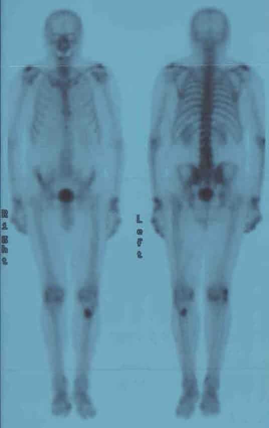
Figure 1. Anterior (right) and posterior (left) view of whole body bone scintigraphy. Increased 99mTc-methylenediphoshonate activity is observed in the proximal diaphysis of the left tibia.
No lesions were visible in the pelvis or in the spine, which made the isolated finding in the tibia unlikely to be related to prostate cancer. The traditional whole body 11C-choline PET/CT, conducted from the cranial basis to the midthigh, did not reveal any pathological uptake site of 11C-choline (Figure 2).
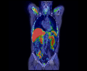
Figure 2. Coronal view of fused 11C-choline PET/CT showing physiological distribution of the tracer. No pathological 11C-choline uptake sites could be detected.
However, since the sensitivity of 11C-choline positron emission tomography/computed tomography (PET/CT) is higher in comparison to bone scintigraphy and also because of the capability of PET/CT to investigate local recurrence and lymph node metastases, the patient underwent subsequent 11C-choline PET/CT to further exclude macroscopic disease. Physiological uptake was seen in the right axillary vein, in the liver, in the spleen, in both kidneys, in the small bowel and in the bladder. In the additional static acquisition centered on the knees, a pathological increase of 11C-choline uptake was seen in the proximal diaphysis of the left tibia which corresponded to the scintigraphic finding (Figure 3A).
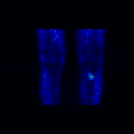
Figure 3A. Coronal view of PET (A) of the inferior limb of at knee height. There is a region of increased uptake of 11C-choline in the area of bone thickening evident in the CT scan.
The CT component of the PET/CT revealed an area of sclerosis (Figure 3B).
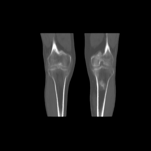
Figure 3B. Coronal view of CT (B, bone window), of the inferior limb at knee height. There is a region of increased uptake of 11C-choline in the area of bone thickening evident in the CT scan.
Fusion imaging demonstrates that the increased metabolic activity localizes to the sclerotic area (Figure 3C).
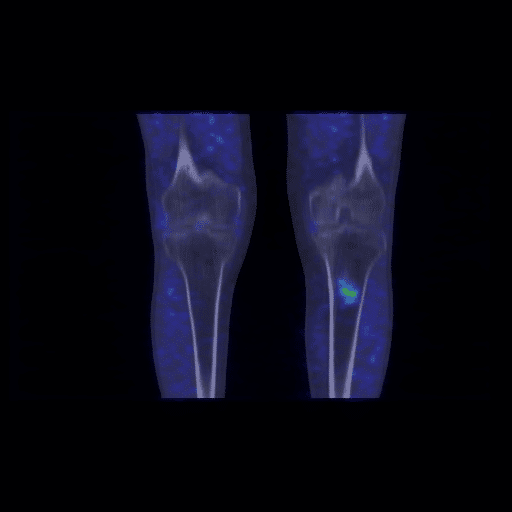
Figure 3C. Coronal view of and fused 11C-choline PET/CT images (C) of the inferior limb at knee height. There is a region of increased uptake of 11C-choline in the area of bone thickening evident in the CT scan.
The patient decided to undergo surgery. Histological analysis revealed enchondroma (Figure 4A and Figure 4B).
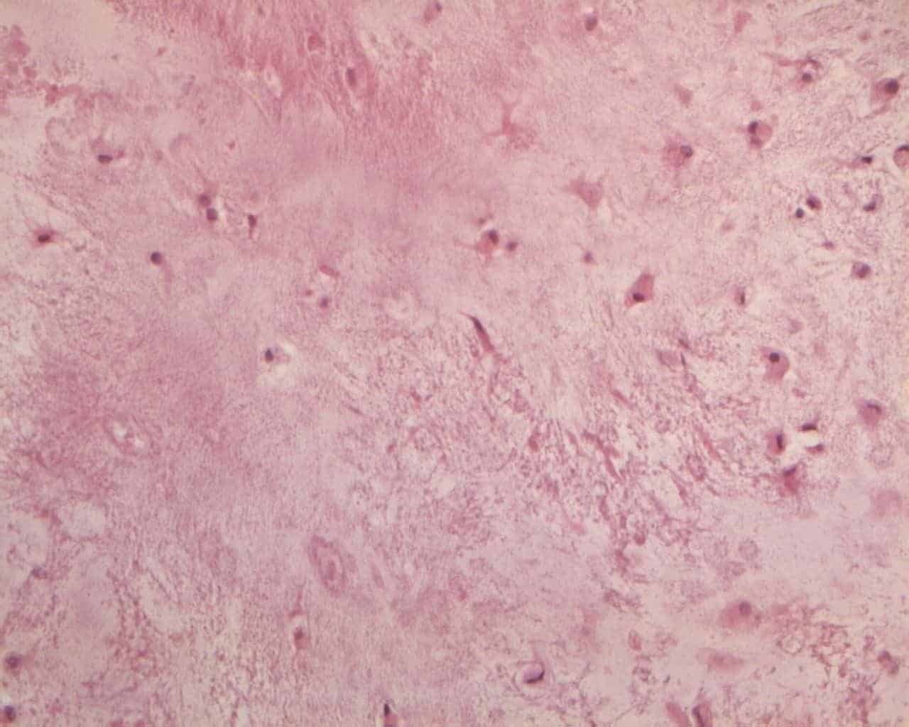
Figure 4A. Typical enchondroma: hypocellular tumor with abundant hyaline cartilage matrix without necrosis and mitosis (e.e. 20x).
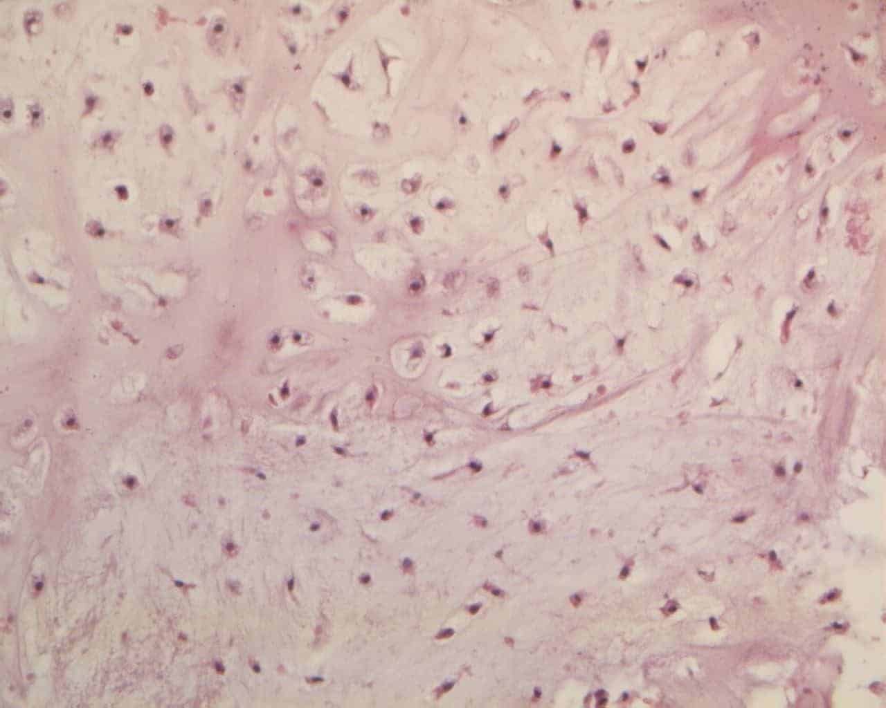
Figure 4B. Chondrocytes with finely granular eosinophilic cytoplasm is often vacuolated with small nuclei (e.e. 20x).
Discussion
Enchondroma is a benign bone tumor, which results from the continued growth of residual benign cartilaginous rests that are displaced from the growth plate. The tumor growth occurs in the medullary cavity of bone [1]. Overall, they are particularly frequent in the phalanges and commonly in the pediatric and young adult age groups, but may also occur in the diaphysis of femur and tibia. This is being recognized in 1.7% of femura at autopsy [2,3]. Enchondromas usually present as a painless bony mass, and radiographically often ovoid in shape with endosteal scalloping; displaying occasionally chondroid type matrix mineralization and do not induce periosteal reactions [4,5]. Bone scintigraphy shows a variable increase of tracer uptake in the skeletal phase whilst the perfusion phase and the blood pool phase are normal [1,2,4]. Malignant transformation into chondrosarcoma is rare but may occur, especially in the diffuse form of multiple enchondromatosis [5,6].
PET/CT with 11C-choline is frequently used for restaging prostate cancer in patients with biochemical failure. Several studies have shown that recurrent disease can be imaged for low PSA values [7–9], that PET/CT with radiolabeled choline might be more accurate than conventional imaging for the detection of lymph node and skeletal metastases [8,10–12], and that several clinical and pathological factors can be used to identify patients who have a higher risk of positive PET/CT [13–15].
Studies indicated that radiolabeled choline accumulates in several malignancies other than prostate cancer or physiological variants, including lung cancer, brain tumors, bladder cancer, meningiomas, as well as inflammatory arthritis disease, Paget’s disease, thymus hyperplasia, benign prostate hyperplasia [16–22]. Increased 11C-choline uptake has also been observed in the pelvic and retroperitoneal lymph nodes of prostate cancer patients with biochemical failure and no histological evidence of disease. This was attributed to lymph node hyperplasia [23]. A simple diffusion mechanism, in addition to an energy-dependent specific transport, regulates the uptake of choline in mammalian cells [24]. Therefore, it is possible that, at least in some of these cases, nonspecific uptake might represent the cause of the false positive.
An alternative hypothesis is that uptake of 11C-choline reflects increased proliferation of cell membranes or of some of their components [25]. It is unknown whether 11C-choline uptake in enchondroma reflects cell proliferation or a concomitant inflammatory process associated with bone remodeling and/or inflammation [26]. The relation between tracer uptake and cellular proliferation is however complex. In humans, the extent of uptake of [11C]choline in the prostate tumor is not related to the cell proliferation rate, as estimated by Ki67 [27].
Nevertheless, in various tumor cells, there was a significant correlation between choline uptake and cell proliferation, as reflected by the incorporation of [3H]methyl-thymidine into DNA [28]. Al-Saeedi et al. found that the concentration of the water-soluble product phosphocholine was higher in breast cancer MCF-7 cells than in control cells [29]. In the same cells, methyl-[3H]choline incorporation was found to be related to the fraction of cells in the S-phase as well as to the incorporation of [methyl-3H]thymidine into DNA [30].
Conclusion
In summary, this case report highlights the necessity of keeping in mind enchondroma in the differential diagnosis of 11C-choline uptake in the skeleton of patients undergoing [11C]choline PET/CT.
Conflict of interest
The authors confirm that this article content has no conflict of interest.
References
- Wang K, Allen L, Fung E, Chan CC, Chan JC, Griffith JF. Bone scintigraphy in common tumors with osteolytic components. Clin Nucl Med. 2005; 30: 655-671.CrossRef PubMed
- Kim MW, Lim ST, Sohn MH. Unusual findings of benign enchondroma in the ulna on 3-phase bone scintigraphy. Clin Nucl Med. 2003; 28: 778-779.CrossRef PubMed
- Scarborough MT, Moreau G. Benign cartilage tumors. Orthop Clin North Am. 1996; 27: 583-589.PubMed
- Murphey MD, Flemming DJ, Boyea SR, Bojescul JA, Sweet DE, Temple HT. Enchondroma versus chondrosarcoma in the appendicular skeleton: differentiating features. Radiographics. 1998; 18: 1213-1237; quiz 44-5.CrossRef PubMed
- Hudson TM, Chew FS, Manaster BJ. Scintigraphy of benign exostoses and exostotic chondrosarcomas. AJR Am J Roentgenol. 1983; 140: 581-586.CrossRef PubMed
- Kenney PJ, Gilula LA, Murphy WA. The use of computed tomography to distinguish osteochondroma and chondrosarcoma. Radiology. 1981; 139: 129-137.CrossRef PubMed
- Castellucci P, Fuccio C, Rubello D, Schiavina R, Santi I, Nanni C, et al. Is there a role for (11)C-choline PET/CT in the early detection of metastatic disease in surgically treated prostate cancer patients with a mild PSA increase <1.5 ng/ml? Eur J Nucl Med Mol Imaging. 2011; 38: 55-63.PubMed
- Giovacchini G, Picchio M, Briganti A, Cozzarini C, Scattoni V, Salonia A, et al. [11C]Choline positron emission tomography/computerized tomography to restage prostate cancer cases with biochemical failure after radical prostatectomy and no disease evidence on conventional imaging. J Urol. 2010; 184: 938-943.CrossRef PubMed
- Giovacchini G, Picchio M, Garcia-Parra R, Mapelli P, Briganti A, Montorsi F, et al. [11C]choline positron emission tomography/computerized tomography for early detection of prostate cancer recurrence in patients with low increasing prostate specific antigen. J Urol. 2013; 189: 105-110.CrossRef PubMed
- Picchio M, Spinapolice EG, Fallanca F, Crivellaro C, Giovacchini G, Gianolli L, et al. [11C]Choline PET/CT detection of bone metastases in patients with PSA progression after primary treatment for prostate cancer: comparison with bone scintigraphy. Eur J Nucl Med Mol Imaging. 2012; 39: 13-26.CrossRef PubMed
- McCarthy M, Siew T, Campbell A, Lenzo N, Spry N, Vivian J, et al. (18)F-Fluoromethylcholine (FCH) PET imaging in patients with castration-resistant prostate cancer: prospective comparison with standard imaging. Eur J Nucl Med Mol Imaging. 2011; 38: 14-22.PubMed
- Beheshti M, Vali R, Waldenberger P, Fitz F, Nader M, Hammer J, et al. The use of F-18 choline PET in the assessment of bone metastases in prostate cancer: correlation with morphological changes on CT. Mol Imaging Biol. 2010; 12: 98-107.CrossRef PubMed
- Castellucci P, Fuccio C, Nanni C, Santi I, Rizzello A, Lodi F, et al. Influence of trigger PSA and PSA kinetics on 11C-Choline PET/CT detection rate in patients with biochemical relapse after radical prostatectomy. J Nucl Med. 2009; 50: 1394-1400.CrossRef PubMed
- Giovacchini G, Picchio M, Coradeschi E, Bettinardi V, Gianolli L, Scattoni V, et al. Predictive factors of [(11)C]choline PET/CT in patients with biochemical failure after radical prostatectomy. Eur J Nucl Med Mol Imaging. 2010; 37: 301-309.PubMed
- Giovacchini G, Picchio M, Scattoni V, Garcia Parra R, Briganti A, Gianolli L, et al. PSA doubling time for prediction of [(11)C]choline PET/CT findings in prostate cancer patients with biochemical failure after radical prostatectomy. Eur J Nucl Med Mol Imaging. 2010; 37: 1106-1116.PubMed
- Farsad M, Schiavina R, Castellucci P, Nanni C, Corti B, Martorana G, et al. Detection and localization of prostate cancer: correlation of (11)C-choline PET/CT with histopathologic step-section analysis. J Nucl Med. 2005; 46: 1642-1649.PubMed
- Fallanca F, Giovacchini G, Ponzoni M, Gianolli L, Ciceri F, Fazio F. Cervical thymic hyperplasia after chemotherapy in an adult patient with Hodgkin lymphoma: a potential cause of false-positivity on [18F]FDG PET/CT scanning. Br J Haematol. 2008; 140: 477.CrossRef PubMed
- Hara T, Inagaki K, Kosaka N, Morita T. Sensitive detection of mediastinal lymph node metastasis of lung cancer with 11C-choline PET. J Nucl Med. 2000; 41: 1507-1513.PubMed
- Roivainen A, Parkkola R, Yli-Kerttula T, Lehikoinen P, Viljanen T, Mottonen T, et al. Use of positron emission tomography with methyl-11C-choline and 2-18F-fluoro-2-deoxy-D-glucose in comparison with magnetic resonance imaging for the assessment of inflammatory proliferation of synovium. Arthritis Rheum. 2003; 48: 3077-3084.CrossRef PubMed
- Kubota K, Furumoto S, Iwata R, Fukuda H, Kawamura K, Ishiwata K. Comparison of 18F-fluoromethylcholine and 2-deoxy-D-glucose in the distribution of tumor and inflammation. Ann Nucl Med. 2006; 20: 527-533.PubMed
- Giovacchini G, Gajate AM, Messa C, Fazio F. Increased C-11 choline uptake in pagetic bone in a patient with coexisting skeletal metastases from prostate cancer. Clin Nucl Med. 2008; 33: 797-798.CrossRef PubMed
- Picchio M, Treiber U, Beer AJ, Metz S, Bossner P, van Randenborgh H, et al. Value of 11C-choline PET and contrast-enhanced CT for staging of bladder cancer: correlation with histopathologic findings. J Nucl Med. 2006; 47: 938-944.PubMed
- Scattoni V, Picchio M, Suardi N, Messa C, Freschi M, Roscigno M, et al. Detection of lymph-node metastases with integrated [11C]choline PET/CT in patients with PSA failure after radical retropubic prostatectomy: results confirmed by open pelvic-retroperitoneal lymphadenectomy. Eur Urol. 2007; 52: 423-429.CrossRef PubMed
- Zeisel SH. Dietary choline: biochemistry, physiology, and pharmacology. Annu Rev Nutr. 1981; 1: 95-121.CrossRef PubMed
- Hara T, Kosaka N, Kishi H. PET imaging of prostate cancer using carbon-11-choline. J Nucl Med. 1998; 39: 990-995.PubMed
- Raisz LG. Physiology and pathophysiology of bone remodeling. Clin Chem. 1999; 45: 1353-1358.PubMed
- Breeuwsma AJ, Pruim J, Jongen MM, Suurmeijer AJ, Vaalburg W, Nijman RJ, et al. In vivo uptake of [11C]choline does not correlate with cell proliferation in human prostate cancer. Eur J Nucl Med Mol Imaging. 2005; 32: 668-673.CrossRef PubMed
- Yoshimoto M, Waki A, Obata A, Furukawa T, Yonekura Y, Fujibayashi Y. Radiolabeled choline as a proliferation marker: comparison with radiolabeled acetate. Nucl Med Biol. 2004; 31: 859-865.CrossRef PubMed
- Al-Saeedi F, Smith T, Welch A. [Methyl-3H]-choline incorporation into MCF-7 cells: correlation with proliferation, choline kinase and phospholipase D assay. Anticancer Res. 2007; 27: 901-906.PubMed
- Al-Saeedi F, Welch AE, Smith TA. [methyl-3H]Choline incorporation into MCF7 tumour cells: correlation with proliferation. Eur J Nucl Med Mol Imaging. 2005; 32: 660-667.CrossRef PubMed
Article information
Corresponding author: Giampiero Giovacchini.
Copyright: © 2016 Giovacchini G, Ciarmiello A.
How to cite: Giovacchini G, Ciarmiello A. Solitary Increase of 11C-Choline Uptake in an Enchondroma Patient with Biochemical Recurrence of Prostate Cancer. J. Diagn. Imaging Ther. 2016; 3(1): 55-58 (https://dx.doi.org/10.17229/jdit.2016-0906-024).
Article history: Received 15 August 2016; Revised 01 September 2016; Accepted 02 September 2016; Published online 06 September 2016.
Archive link: JDIT-2016-0906-024
You are here: home »
