Keynote: A high-resolution MRI ensemble model combining plaque and perivascular adipose tissue radiomics can match PET in detecting carotid plaque inflammation and predicting disease progression. This approach offers a radiation-free alternative for identifying high-risk patients with asymptomatic carotid atherosclerosis.
Keywords: MRI radiomics, carotid atherosclerosis, perivascular adipose tissue, plaque inflammation, PET-equivalent imaging, stroke prevention
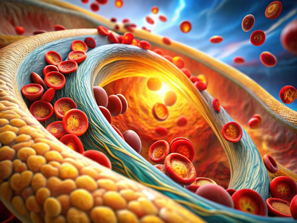
Carotid atherosclerosis is a major contributor to strokes and other cerebrovascular events worldwide. The risk of severe events often stems from inflammation-driven progression and rupture of plaque. Traditionally, 18F-fluorodeoxyglucose ([18F]FDG) PET has been the gold standard for identifying inflammation in carotid plaques; however, its use is limited by concerns regarding radiation exposure and cost.
A new prospective study published in EJNMMI Research demonstrates that a novel MRI-based ensemble model, which combines radiomics data from carotid plaque and surrounding perivascular adipose tissue (PVAT), can match PET performance in detecting highly inflammatory plaques (HIPs) and predicting plaque progression. This finding opens the door to safer, more accessible screening for patients at risk of cerebrovascular events.
Why Inflammation Matters in Carotid Plaques
Inflammatory activity within carotid plaques increases their vulnerability, making rupture more likely. This inflammation is often driven by macrophage activity, which destabilises plaque structure through the release of enzymes like matrix metalloproteinases (MMPs).
While PET is effective in quantifying inflammation through glucose uptake (SUVmax), it is not suitable for widespread screening due to the radiation dose and limited availability. High-resolution vessel wall MRI (HR-VW MRI) offers a non-invasive, radiation-free alternative — and by applying radiomics, researchers can extract detailed quantitative features that reflect tissue heterogeneity linked to inflammation.
Study Overview
The research team at Xuanwu Hospital, China, enrolled 159 patients with asymptomatic carotid atherosclerosis between April 2022 and August 2023. All participants underwent integrated PET/MRI scanning, enabling direct comparison between MRI radiomics and PET findings.
Key features of the study design:
- 209 carotid plaques analysed (104 HIPs)
- Both plaque and PVAT were characterised using radiomics (3,864 features each)
- Patients were followed for 12 months with a repeat MRI to assess plaque progression
- An ensemble model built by combining plaque and PVAT radiomics scores, compared against the PET SUVmax performance
What the Researchers Found
Distinct MRI Features in Highly Inflammatory Plaques
HIPs had larger lipid cores, more intraplaque haemorrhage (IPH), and less calcification than non-HIP plaques. They also showed a higher prevalence of indistinct PVAT (IPVAT) — a sign of altered fat tissue adjacent to inflamed vessels — in both training and testing datasets.
Strong PVAT–Inflammation Link
IPVAT consistently correlated with PET SUVmax in both HIPs and non-HIPs, suggesting PVAT changes mirror inflammatory processes in the plaque itself. This reinforces the role of PVAT as a potential biomarker for vascular inflammation.
Ensemble Model Performance
The combined plaque + PVAT radiomics model achieved:
- AUC 0.92 (training) and 0.91 (testing) for identifying HIPs
- Equal accuracy to PET (AUC 0.79 vs. 0.85) in predicting plaque progression at 12 months
- A 16–20% improvement in predictive accuracy compared to plaque- or PVAT-only models
Clinical Implications
By integrating multiple MRI-derived features, the ensemble model effectively creates a “virtual PET” image without the need for radioactive tracers. This could make routine assessment of carotid plaque inflammation feasible in more centres, especially where PET is not available or practical.
The ability to identify high-risk plaques early could allow clinicians to:
- Target anti-inflammatory therapies more precisely
- Monitor plaque stability over time
- Reduce unnecessary PET scans in low-risk cases
Limitations and Next Steps
The study was conducted at a single centre, so multicentre validation is needed to confirm generalisability. Longer-term follow-up is also necessary to assess whether the model can accurately predict stroke risk, not just plaque progression.
Future research may focus on improving scanner-to-scanner reproducibility and integrating this method into clinical decision pathways for vascular disease.
Take-Home Message
This study demonstrates that MRI-based radiomics, when applied to both plaque and PVAT, can match the performance of PET in detecting inflammation and predicting the progression of carotid atherosclerosis. The approach offers a promising, radiation-free tool for identifying patients at the highest risk — and could become an important step in preventing stroke.
Reference: Yu, F., Li, X., Zhang, Y. et al. MRI ensemble model of plaque and perivascular adipose tissue as PET-equivalent for identifying carotid atherosclerotic inflammation. EJNMMI Res 15, 103 (2025). https://doi.org/10.1186/s13550-025-01293-9
Disclaimer: This article summarises and interprets peer-reviewed research for educational purposes. It is not a substitute for professional medical advice. Always consult a qualified healthcare provider for clinical decision-making.