PET imaging represents a pinnacle in modern medical diagnostics, offering unparalleled insights into the body’s metabolic processes. This article explores the mechanism of PET imaging, its clinical applications, the procedure, and the future of this revolutionary technology in healthcare. With its ability to detect diseases at their onset and monitor treatment efficacy, PET imaging stands as a vital tool in contemporary medicine.
Overview of PET Imaging
Positron Emission Tomography (PET) imaging is a revolutionary medical modality technique that has transformed the way diseases, particularly cancers, neurological disorders, and cardiac conditions, are diagnosed and treated. Unlike traditional imaging methods such as X-rays or MRI, PET scans provide a dynamic picture of what’s happening inside the body at a molecular level. This detailed insight allows physicians to see how organs and tissues are functioning in real-time, making it an invaluable tool in modern medicine.
Understanding the Basics of PET
PET imaging is based on the detection of gamma rays emitted from a radioactive substance administered to the patient. This substance, known as a radiotracer, is typically a biologically active molecule labelled with a radioactive isotope, such as glucose. The most commonly used tracer in PET imaging is fluorodeoxyglucose (FDG), a glucose analogue. When injected into the body, FDG travels through the bloodstream and accumulates in cells that use glucose for energy.
The Mechanism of PET Imaging
The mechanism of PET imaging hinges on the metabolic activity of cells. These cells in the body, particularly those growing rapidly, consume glucose as a source of energy. Cancer cells, for instance, are known to metabolise glucose much faster than normal cells. When FDG is injected, it gets taken up by these active cells, but unlike normal glucose, it does not undergo further metabolism. This accumulation of FDG makes rapidly growing cells, like cancer cells, visible on a PET scan.
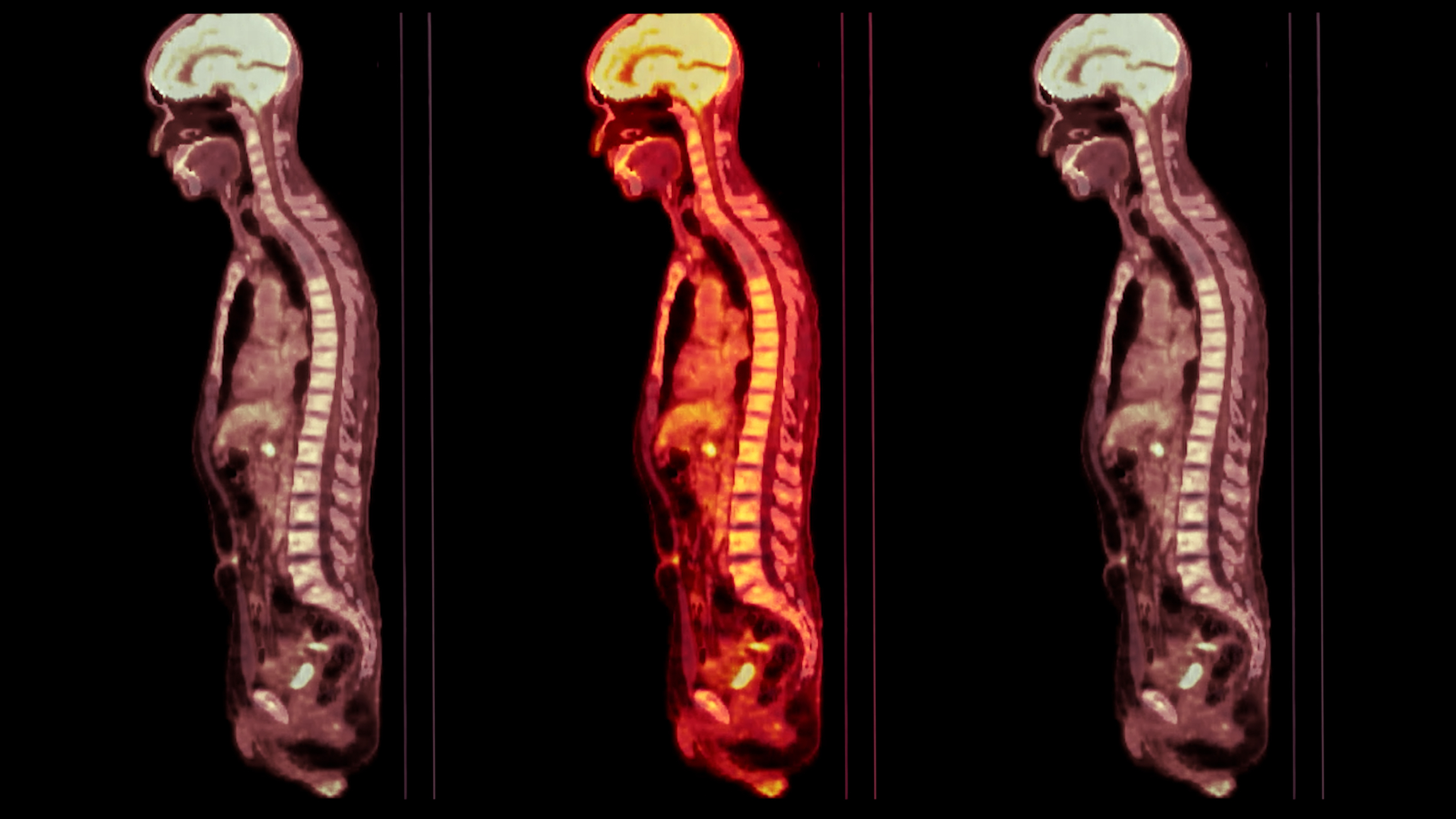
When the FDG decays, it emits positrons, the antimatter counterparts of electrons. Upon encountering electrons, these positrons undergo annihilation, leading to the emission of gamma rays. The PET scanner then detects these rays, which circle around the patient, capturing images from multiple angles. A computer then reconstructs these images into detailed, three-dimensional representations of the patient’s internal metabolic processes.
The Procedure of a PET Scan
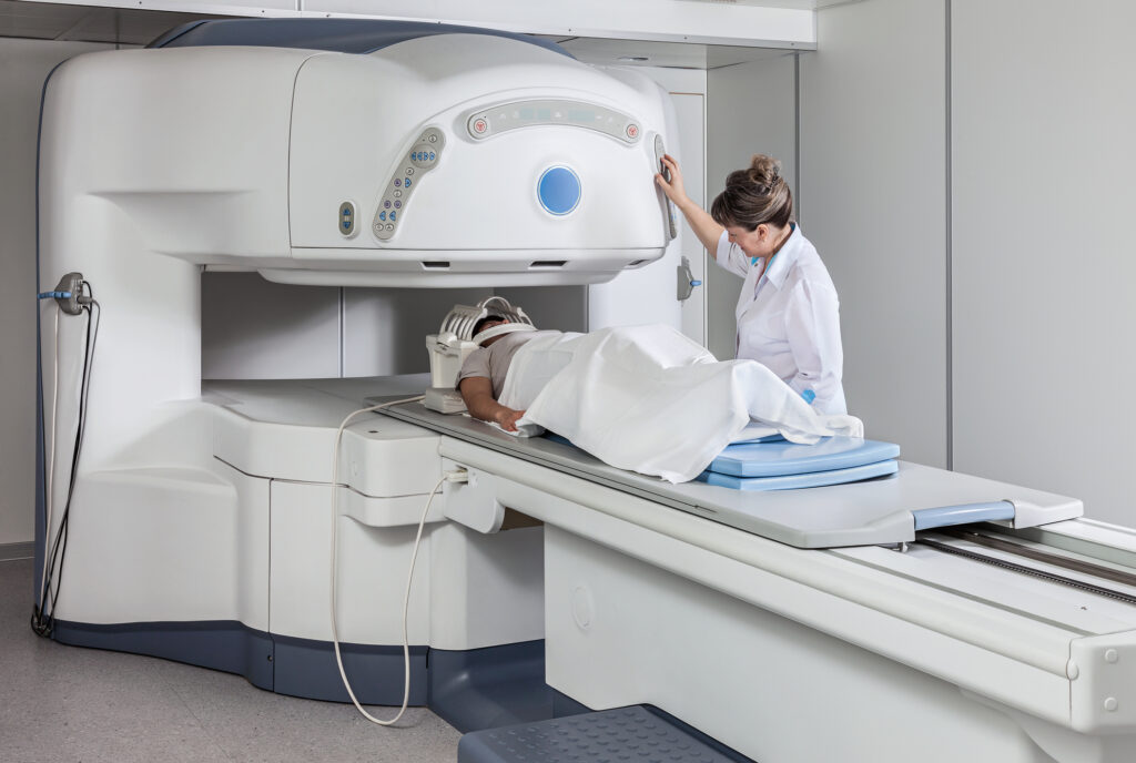
A PET scan is a relatively straightforward but highly sophisticated procedure. Before the scan, the patient is injected with the FDG tracer. A waiting period, typically around an hour, allows the tracer to distribute and accumulate in the body. The patient is then placed in the PET scanner, which resembles a large donut-shaped machine.
During the scan, it is crucial for the patient to remain as still as possible to ensure clear images. The scanning process usually takes about 30 to 45 minutes, depending on the examined area. The PET scanner detects the gamma rays emitted from the patient’s body and sends this data to a computer, which constructs it into images for analysis.
Advantages of PET Imaging Over Other Techniques
The major advantage of PET over other diagnostic methods lies in its ability to detect changes at the cellular level, often before structural changes become apparent on X-rays or MRI. This early detection is crucial, especially in cancer treatment, where early intervention can significantly improve prognosis.
PET scans are also incredibly useful in assessing the effectiveness of treatments. By comparing scans taken before and after treatment, doctors can determine if a tumour is responding to therapy, providing critical feedback that can guide further treatment decisions.
Applications of PET Imaging
While PET is most prominently used in oncology for detecting and monitoring cancer, it has a wide array of other applications. In cardiology, PET scans are used to assess heart function, particularly in cases of coronary artery disease, to evaluate the extent of damage after a heart attack. In neurology, PET plays a crucial role in diagnosing conditions such as Alzheimer’s disease, epilepsy, and other brain disorders. It can highlight areas of the brain that are overactive or underactive, providing essential clues to the underlying pathology.
Safety and Considerations
PET imaging is generally safe, with minimal risks involved. The amount of radiation from the tracer is low and typically clears from the body within a few hours. However, as with any procedure involving radiation, it is not usually recommended for pregnant women or those who are breastfeeding. Additionally, patients with diabetes may require special preparation due to the involvement of the glucose-based tracer.
PET imaging stands as a beacon of innovation in diagnostic imaging. By offering insights into the body’s metabolic processes, it opens new avenues in the early detection and treatment of various diseases. As technology progresses, the scope and accuracy of PET are expected to advance further, underpinning its essential role in the future of healthcare diagnostics.
Clinical Applications of PET Imaging
Positron Emission Tomography (PET) imaging is a highly advanced diagnostic tool that has significantly impacted the field of medical imaging. Its unique ability to visualise metabolic processes in the body has made it an indispensable tool in several key areas of medicine, including oncology, cardiology, and neurology. This article explores the diverse clinical applications of PET imaging, highlighting its role in diagnosis, staging, and treatment monitoring across various medical specialties.
Oncology: A Cornerstone of PET Imaging
In oncology, PET plays a pivotal role in the management of cancer patients. It is particularly effective in diagnosing, staging, and monitoring the treatment response of various cancers.
- Diagnosis: PET scans can detect the presence of cancer by revealing the metabolic activity of cells. Cancer cells typically have higher metabolic rates than normal cells, making them easily identifiable on PET scans.
- Staging: Determining the stage of cancer is crucial for treatment planning. PET imaging provides comprehensive information about the extent of the disease, including the presence of metastases (spread of cancer to other parts of the body), which other imaging modalities might not detect.
- Treatment Monitoring: PET scans are invaluable in assessing the effectiveness of cancer treatments, such as chemotherapy or radiation therapy. Changes in the metabolic activity of a tumour, as seen on a PET scan, can indicate how well the tumour is responding to treatment, often before physical changes are detectable.
- Recurrence Detection: PET imaging is also used to detect the recurrence of cancer, helping to initiate timely interventions.
Cardiology: Assessing Heart Function
In cardiology, PET imaging has become a critical tool for assessing heart function and guiding treatment strategies.
- Myocardial Perfusion Imaging: PET is used to evaluate blood flow to the heart muscle (myocardial perfusion). This is particularly important in diagnosing coronary artery disease and determining the extent of damage caused by a heart attack.
- Cardiac Viability: PET scans can also assess the viability of heart tissue, especially in patients who have had previous heart attacks. This helps in identifying areas of the heart that are still functional and can respond to treatment.
- Guiding Treatment Decisions: The detailed information provided by PET imaging assists cardiologists in making informed decisions about treatments such as angioplasty, bypass surgery, or medication management.
Neurology: Exploring Brain Function
PET imaging has made significant contributions to the field of neurology, particularly in diagnosing and managing neurological disorders.
- Alzheimer’s Disease: PET scans are used to detect the early signs of Alzheimer’s disease by identifying patterns of brain activity and amyloid plaque deposits, which are hallmarks of the disease.
- Epilepsy: In epilepsy, PET imaging is used to localise the area of the brain where seizures originate, aiding in treatment planning, especially in cases where surgery is considered.
- Brain Tumours: PET helps differentiate between benign and malignant brain tumours and assess the response to therapy.
- Other Neurological Disorders: PET scans are also valuable in diagnosing and managing other disorders such as Parkinson’s disease, Huntington’s disease, and stroke.
Enhancing Diagnostic Accuracy with Hybrid Imaging
PET scans are often combined with CT (Computed Tomography) or MRI (Magnetic Resonance Imaging) scans in a technique known as hybrid imaging. This combination provides both metabolic and anatomical information, significantly enhancing the accuracy of diagnosis and treatment planning. For instance, a PET/CT scan combines the detailed metabolic information from a PET scan with the high-resolution anatomical images from a CT scan, providing a more comprehensive view of the patient’s condition.
The PET Imaging Procedure
The PET imaging procedure is both non-invasive and painless, offering a patient-friendly approach to diagnostic imaging.
- Preparation and Tracer Injection: Patients are usually required to fast for a certain period before the test. A radioactive tracer, typically FDG, is then injected into the bloodstream.
- Resting Period: After the injection, there is a resting period, usually about an hour, to allow the tracer to distribute throughout the body.
- Scanning Process: The actual scanning process takes about 30 minutes. During this time, the patient lies on a table that slides into the PET scanner.
- Safety and Radiation Exposure: The procedure is safe, with the radiation exposure from the tracer being relatively low and comparable to that received during a standard X-ray.
PET imaging has revolutionised the field of diagnostic imaging, providing critical insights into various diseases. Its applications in oncology, cardiology, and neurology have notably enhanced the accuracy of diagnoses, efficacy of treatment plans, and overall patient care. As technology advances, the scope and capabilities of PET imaging are expected to expand further, cementing its role as a key component in modern medicine.
Advancements in PET Imaging Technology
Positron Emission Tomography (PET) imaging, an essential tool in modern medical diagnostics, has witnessed significant advancements over recent years. These developments enhance PET scans’ quality and efficiency and broaden their applications in personalised medicine. This article explores the latest advancements in PET imaging technology, focusing on improvements in scan resolution and speed, the integration of digital and AI technologies, and the development of novel tracers.
Enhanced Resolution and Speed
The latest PET scanners boast considerably improved resolution and faster scanning times. This progress is primarily due to advancements in detector technology. Modern PET scanners use smaller, more sensitive detectors that provide higher-resolution images. This improvement allows for the detection of smaller lesions, which is critical in early-stage cancer diagnosis. Additionally, faster scan times reduce patient discomfort and inconvenience and increase the throughput of scanning facilities.
Digital PET Scanners
The evolution from analogue to digital PET scanners marks a significant technological leap. Digital PET scanners, equipped with solid-state silicon photomultipliers, offer superior sensitivity and improved time-of-flight (TOF) measurements compared to traditional photomultiplier tubes. This upgrade results in sharper images with better contrast, aiding in more accurate disease characterisation. The digital technology also reduces noise and enhances signal processing, further improving image quality.
Integration of AI Algorithms
Artificial Intelligence (AI) plays a pivotal role in the evolution of PET imaging. AI algorithms are being integrated into PET imaging to improve image reconstruction, reduce artefacts, and enhance overall image quality. These algorithms can also analyse vast amounts of imaging data quickly, aiding in the identification of subtle patterns that the human eye might miss. This capability is particularly beneficial in complex cases where precise diagnosis is crucial.
Development of Novel Tracers
One of the most exciting areas of advancement in PET imaging is the development of novel tracers. Traditionally, FDG has been the mainstay tracer used in PET scans. However, researchers are now developing a variety of new tracers designed to target specific diseases. These tracers can bind to particular proteins or cells associated with certain types of cancer or neurological disorders, allowing for more targeted and accurate diagnosis. This advancement is particularly promising in the sphere of personalised medicine, where treatment can be tailored to the individual characteristics of a patient’s disease.
PET/MRI Hybrid Imaging
The combination of PET with Magnetic Resonance Imaging (MRI) in hybrid scanners is another significant advancement. PET/MRI offers the metabolic and functional imaging capabilities of PET alongside the superior soft tissue contrast of MRI. This combination is particularly useful in neurological and oncological applications, where detailed anatomical information is crucial. Additionally, PET/MRI exposes patients to less radiation compared to PET/CT, making it a safer option, especially for children and patients requiring multiple scans.
Quantitative PET Imaging
Advancements in quantitative PET imaging enable the measurement of absolute levels of tracer uptake in tissues. This quantification is essential for accurately assessing treatment response and disease progression. Quantitative PET imaging is becoming increasingly important in clinical trials, where precise measurements of how a drug affects a tumour are crucial.
Safety and Considerations
Despite these advancements, safety considerations remain paramount in PET imaging. The use of radioactive tracers necessitates careful handling and disposal procedures to minimise radiation exposure to patients and healthcare workers. While the radiation dose from a single PET scan is generally low, cumulative exposure from multiple scans can be a concern.
Special Considerations
- Pregnancy and Breastfeeding: Pregnant or breastfeeding women should consult their doctor before undergoing a PET scan, as the tracer could potentially affect the fetus or infant.
- Diabetic Patients: Special preparation is required for diabetic patients due to the involvement of glucose-based tracers. Blood sugar levels need to be carefully managed before the scan.
- Allergic Reactions: Although rare, allergic reactions to the tracer can occur. Patients should inform their healthcare provider of any known allergies.
The advancements in PET imaging technology represent a quantum leap in medical diagnostics. With enhanced resolution, speed, and the development of novel tracers, PET imaging is becoming more precise and personalised. The integration of digital and AI technologies further amplifies its capabilities, paving the way for more accurate and early diagnosis of diseases. As the technology continues to evolve, PET imaging is set to play an even more critical role in the future of healthcare, offering new opportunities for improved patient outcomes while maintaining a strong focus on safety and efficacy.
The Role of PET Imaging in Personalised Medicine
Personalised medicine, a rapidly evolving approach in healthcare, aims to tailor treatment to the individual characteristics of each patient. Positron Emission Tomography (PET) imaging has emerged as a crucial tool in this paradigm, offering detailed insights at the molecular and cellular levels. This article discusses the integral role of PET imaging in personalised medicine, highlighting its impact on treatment efficacy and patient care while also addressing the challenges and future directions of this technology.
Personalised Diagnosis and Treatment Planning
PET imaging’s ability to provide detailed metabolic and physiological information makes it invaluable in creating personalised treatment plans. By identifying specific characteristics of diseases, particularly cancer, PET imaging allows clinicians to choose the most effective treatment strategies for individual patients.
- Cancer Treatment: PET scans can reveal tumours’ metabolic activity and aggressiveness, guiding oncologists in selecting the most appropriate treatment. For instance, in cases where a tumour demonstrates high metabolic activity on a PET scan, more aggressive treatment may be warranted.
- Assessing Treatment Response: PET imaging is highly effective in monitoring how a tumour responds to treatment. Changes in the metabolic activity of a tumour, as seen on a PET scan, can indicate the effectiveness of a particular therapy, allowing for adjustments in treatment plans.
- Avoiding Unnecessary Treatments: By accurately assessing the extent of disease and treatment response, PET imaging helps avoid ineffective treatments, thereby sparing patients from unnecessary side effects and procedures.
Tailoring Treatment to Genetic Profiles
The integration of PET imaging with genetic and molecular analysis is a significant step forward in personalised medicine. PET scans can be used in conjunction with genetic testing to identify specific mutations or markers in tumours, guiding the selection of targeted therapies.
- Targeted Radiopharmaceuticals: The development of novel PET tracers that target specific molecular pathways or receptors in cancer cells is underway. These tracers can illuminate specific biological processes, enabling more precise treatment.
- Pharmacodynamics: PET imaging helps in understanding the pharmacodynamics of drugs, showing how a drug is distributed and metabolised in the body. This information is crucial for determining each patient’s optimal drug and dosage.
Enhancing Drug Development
PET imaging is increasingly used in clinical trials to assess the efficacy of new drugs. By providing real-time feedback on how a drug affects a disease process at the molecular level, PET imaging accelerates the development of effective therapies and helps in making more informed decisions during the drug development process.
Overcoming Challenges in PET Imaging
Although its potential, PET imaging faces several challenges to fully realise its role in personalised medicine.
- High Costs: The high cost of PET scans, largely due to expensive tracers and sophisticated equipment, limits widespread accessibility. Efforts are underway to develop more cost-effective PET imaging methods.
- Limited Availability of Tracers: Currently, the range of available tracers is limited, restricting the scope of diseases that can be effectively studied using PET. Research is focused on developing a broader spectrum of tracers to target various diseases.
- Need for Specialised Equipment and Personnel: PET imaging requires specialised equipment and trained personnel, which are not available in all healthcare settings. This limitation poses a challenge in integrating PET imaging into routine clinical practice.
Future Directions in PET Imaging
The future of PET imaging in personalised medicine is promising, with several advancements on the horizon.
- Integration with Other Imaging Modalities: Combining PET with other imaging techniques like MRI and CT can provide comprehensive diagnostic information, enhancing the accuracy of personalised treatment plans.
- Artificial Intelligence in PET Imaging: The use of AI in interpreting PET images can improve diagnostic accuracy and efficiency. AI algorithms can analyse complex imaging data to identify patterns and make predictive analyses.
- Expanding Tracer Development: Ongoing research in radiopharmaceuticals is focused on creating novel tracers that can target a wider range of diseases and biological processes. This development is crucial for expanding the role of PET in personalised medicine.
- Reducing Radiation Exposure: Efforts to reduce the radiation dose associated with PET imaging without compromising image quality are important, especially for patients requiring multiple scans.
PET imaging stands at the forefront of the personalised medicine revolution, offering unmatched insights into the molecular workings of diseases. Its ability to guide tailored treatment plans, monitor treatment efficacy, and assist in drug development solidifies its position as an invaluable tool in modern healthcare. While challenges such as cost and availability of tracers persist, ongoing advancements in technology and tracer development promise to overcome these hurdles, paving the way for wider application and integration of PET imaging in personalised medicine. As this field evolves, it holds the potential to significantly improve patient outcomes by providing more targeted, effective, and efficient healthcare.
Conclusion
Positron Emission Tomography (PET) imaging stands as a pivotal advancement in the sphere of medical diagnostics. This sophisticated technology transcends traditional diagnostic methods by providing a window into the body’s metabolic and molecular processes, offering invaluable insights that are crucial for effective disease management. As we have explored, PET imaging’s applications span a broad spectrum of medical fields, from oncology and cardiology to neurology, each benefiting from its ability to deliver precise and personalised diagnostic information.
The integration of PET imaging into the clinical setting has revolutionised the approach to disease diagnosis and treatment. It has become instrumental in early cancer detection, staging, and monitoring treatment responses in oncology, allowing for more targeted and effective therapies. In cardiology, PET imaging provides essential data on myocardial perfusion and viability, guiding treatment decisions for heart disease. Furthermore, neurology aids in understanding complex brain functions and disorders, offering crucial information for managing conditions like Alzheimer’s disease and epilepsy.
The advancements in PET technology, such as the development of digital PET scanners, the integration of AI algorithms, and the creation of novel tracers, continue to enhance its diagnostic capabilities. These innovations improve the resolution and speed of scans and pave the way for more personalised treatment approaches. The evolution of PET imaging aligns closely with personalised medicine principles, where treatments are increasingly tailored to individual patient profiles, maximising efficacy while minimising unnecessary side effects.
Notwithstanding challenges like high costs and the need for specialised equipment and personnel, the future of PET imaging is bright. As research progresses, it promises to expand its role in healthcare, offering more accessible and diverse applications. PET imaging is not just a tool for today’s medical professionals; it is a beacon for future medical innovations, continually reshaping our understanding of disease and revolutionising patient care in the process. With its profound impact on healthcare, this technology reaffirms its status as an indispensable asset in modern medicine, poised to enhance patient outcomes and advance the field for years to come.
Disclaimer
The content provided in this article is for informational and educational purposes only and does not constitute medical advice, diagnosis, or treatment. While every effort has been made to ensure the accuracy and currency of the information presented, Open Medscience does not guarantee its completeness or reliability. Readers should consult qualified healthcare professionals for advice tailored to their specific medical needs or conditions. The technologies and techniques described, including Positron Emission Tomography (PET) imaging, may not be suitable or available in all clinical settings, and individual patient outcomes may vary. Open Medscience disclaims any liability for decisions made based on the information presented in this article.
home » blog » medical imaging topics »