Bone health is integral to overall well-being, particularly as the person ages. Osteoporosis, a condition characterised by decreased bone density and increased fracture risk, affects millions globally. Early detection and management are paramount in mitigating its impact. Bone densitometry, specifically DEXA, plays a pivotal role in this regard. This article explores the intricacies of DEXA, elucidating its significance, methodology, and clinical applications.
What is Bone Densitometry?
Bone densitometry, or DEXA, is a specialised form of X-ray technology that measures bone mineral density (BMD). It utilises two X-ray beams with different energy levels to produce detailed images of the bone. By comparing the absorption of each beam by the bone, the scanner calculates the bone density. The lower the bone density, the higher the risk of fractures.
The Science Behind DEXA
DEXA operates on the principle of differential absorption. Bones, being denser than soft tissues, absorb more X-rays. The DEXA machine emits two X-ray beams at different energy levels. As these beams pass through the body, they are absorbed by bones and soft tissues at different rates. The amount of X-ray that passes through is detected and measured, allowing the calculation of bone mineral density.
Measurement Sites
Common sites for DEXA scans include the lumbar spine, hip, and forearm. These areas are most prone to osteoporotic fractures. DEXA provides valuable insights into bone health and fracture risk by focusing on these critical regions.
Clinical Applications of DEXA
Osteoporosis is often termed a “silent disease” as it progresses without symptoms until a fracture occurs. DEXA is the gold standard for diagnosing osteoporosis. It quantifies BMD and compares it with reference values, typically young adult mean BMD. A T-score is derived from this comparison:
- Normal: T-score above -1
- Osteopenia: T-score between -1 and -2.5
- Osteoporosis: T-score below -2.5
Assessing Fracture Risk
Beyond diagnosing osteoporosis, DEXA assesses fracture risk. Low BMD is a significant predictor of fractures. Coupled with clinical risk factors (age, gender, history of fractures), DEXA helps formulate a comprehensive risk profile. This information is crucial for preventive strategies and treatment planning.
Monitoring Treatment Efficacy
Patients undergoing treatment for osteoporosis or other bone-related conditions require regular monitoring to gauge the effectiveness of interventions. DEXA scans are periodically used to track changes in BMD. An increase in BMD suggests a positive response to treatment, while a decrease might necessitate a review of the therapeutic approach.
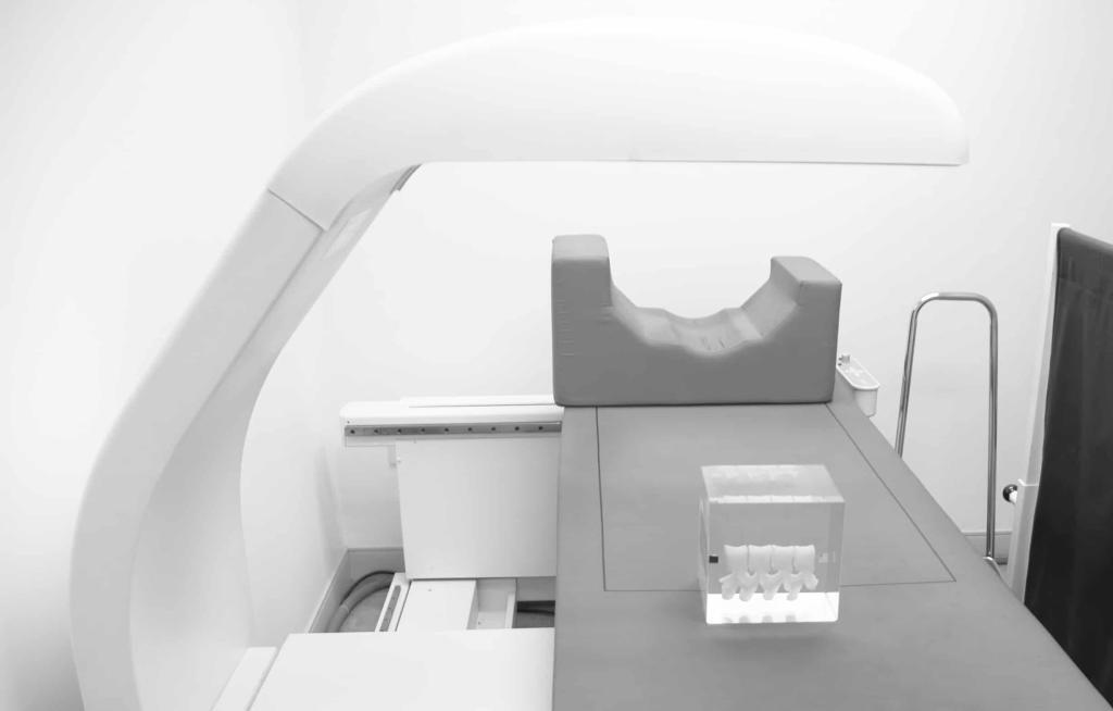
The DEXA Procedure
Preparation for a DEXA scan is minimal. Patients are advised to avoid calcium supplements for 24 hours before the test. Comfortable clothing without metal zippers or buttons is recommended. The procedure is quick, painless, and typically completed within 20 minutes.
The Scanning Process
- Positioning: The patient lies on a padded table. The area of interest (spine, hip, or forearm) is positioned under the DEXA scanner.
- Scanning: The machine passes a small arm over the area, emitting X-ray beams. The patient remains still during the scan.
- Image Acquisition: The machine captures images and data, which are then processed to calculate BMD.
Safety Considerations
DEXA is a safe procedure with minimal radiation exposure. The radiation dose from a DEXA scan is significantly lower than that of a standard chest X-ray. As with any medical procedure, pregnant women should inform their healthcare provider to assess the necessity and safety of the scan.
Interpretation of DEXA Results
- T-Score: Compares the patient’s BMD with the mean BMD of a healthy young adult. It is used to diagnose osteoporosis.
- Z-Score: Compares the patient’s BMD with the mean BMD of people in the same age group, gender, and ethnicity. It helps identify whether factors other than ageing are contributing to bone loss.
Clinical Implications
A T-score below -2.5 confirms osteoporosis, necessitating interventions to prevent fractures. Lifestyle modifications and preventive treatments may be recommended for those with osteopenia. Regular follow-up scans help assess the progression of bone density loss and the effectiveness of treatments.
Advances in Bone Densitometry
Technological advancements have enhanced the accuracy and precision of DEXA scans. Modern DEXA machines offer better image resolution, faster scanning times, and more detailed assessments of bone health. Some machines now provide additional data on body composition, including fat and lean mass.
Peripheral DEXA (pDEXA)
Peripheral DEXA (pDEXA) scans focus on peripheral sites like the forearm, heel, and finger. These scans are less common but useful in specific situations where central DEXA is not feasible. Due to its portability and lower cost, pDEXA is often used in large-scale screening programmes.
Vertebral Fracture Assessment (VFA)
Vertebral fractures are a common consequence of osteoporosis. VFA, often performed alongside DEXA, uses similar technology to detect fractures in the spine. This combined approach enhances the diagnostic capability, providing a comprehensive evaluation of bone health.
Benefits of DEXA
Early detection of low bone density allows for timely intervention, reducing the risk of fractures. Lifestyle changes, dietary modifications, and medications can significantly improve bone health and quality of life.
Non-Invasive and Quick
DEXA is non-invasive, painless, and requires no special preparation. The quick nature of the scan makes it convenient for patients, ensuring high compliance rates.
Comprehensive Risk Assessment
DEXA thoroughly assesses fracture risk by combining BMD measurements with clinical risk factors. This holistic approach aids in personalised treatment plans tailored to individual needs.
Limitations of DEXA
DEXA primarily measures bone density and does not provide information on bone quality or microarchitecture. For a more detailed assessment, other imaging techniques, like quantitative computed tomography (QCT), may be needed.
Radiation Exposure
Though minimal, DEXA does involve exposure to ionising radiation. While the dose is low, it is still a consideration, particularly for patients requiring frequent scans.
Accessibility and Cost
DEXA machines are not universally available, particularly in low-resource settings. The cost of the scan may also be a barrier for some patients, limiting access to this valuable diagnostic tool.
Future Directions in Bone Densitometry
Future advancements may see DEXA integrated with other imaging modalities for a more comprehensive assessment of bone health. Combining DEXA with technologies like MRI or QCT could provide insights into bone quality and strength beyond density measurements.
Improved Predictive Models
Research is ongoing to refine predictive models that combine DEXA results with genetic, biochemical, and clinical data. These models aim to enhance the accuracy of fracture risk predictions, leading to better-targeted interventions.
Portable and Accessible Solutions
Innovations in portable DEXA machines and cost-effective solutions are on the horizon. These advancements aim to improve accessibility, particularly in underserved regions, ensuring that more people can benefit from early osteoporosis detection and management.
Conclusion
Bone densitometry, specifically DEXA, is a cornerstone in the diagnosis and management of osteoporosis. Its ability to accurately measure bone mineral density and assess fracture risk makes it an invaluable tool in modern medicine. Understanding DEXA’s principles, applications, and limitations empowers healthcare providers and patients to make informed decisions about bone health. As technology advances, the future of bone densitometry promises even greater accuracy, accessibility, and integration, enhancing our ability to combat osteoporosis and improve quality of life.
This article underscores the significance of bone densitometry in preventive healthcare by providing a detailed overview of DEXA. From early detection to ongoing monitoring, DEXA stands as a testament to the strides made in medical imaging and its impact on patient outcomes.
Disclaimer
The information provided in this article is intended for educational and informational purposes only. It is not a substitute for professional medical advice, diagnosis, or treatment. While efforts have been made to ensure accuracy, the content should not be relied upon as a basis for making health-related decisions without consulting a qualified healthcare professional. Bone densitometry, including DEXA scans, should be conducted and interpreted by trained medical practitioners in the context of an individual’s full clinical history. Readers are advised to speak to their GP, specialist, or other healthcare provider regarding any medical concerns or before beginning any treatment related to bone health or osteoporosis. The authors and publishers disclaim any liability for adverse outcomes resulting from the use or application of the information contained herein.
home »