Mild traumatic brain injury (mTBI), or concussion, is one of the most prevalent neurological conditions globally. It is frequently caused by contact sports injuries, accidental falls, road traffic accidents, and blast-related or blunt-force trauma in military contexts. While labelled “mild” because it does not typically involve extended loss of consciousness or large visible lesions on imaging, the functional impact can be considerable.
Many patients experience a combination of physical, cognitive, and emotional symptoms. Common complaints include headaches, dizziness, fatigue, poor concentration, memory difficulties, irritability, and disturbances in sleep. In some individuals, these symptoms resolve within days or weeks; in others, they can persist for months or even years.
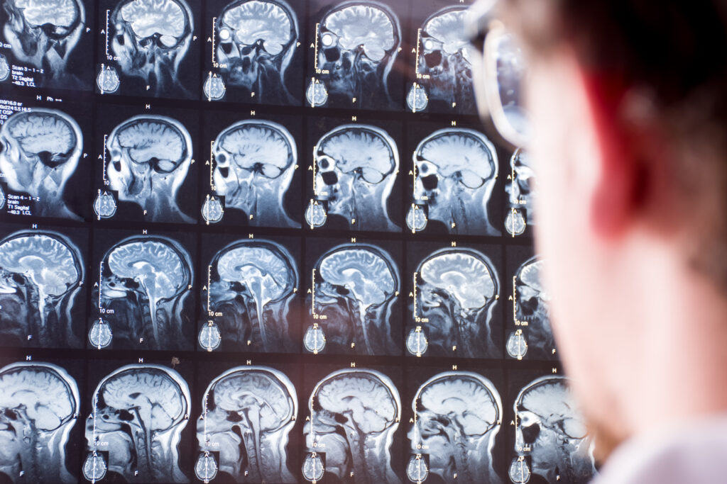
From a diagnostic perspective, one of the most significant challenges with mTBI is that conventional imaging techniques — such as computed tomography (CT) or standard MRI sequences — often appear entirely normal. The absence of visible abnormalities on scans can make it difficult to validate the patient’s symptoms, complicate return-to-activity decisions, and leave uncertainty in both clinical and legal contexts.
Over the past decade, researchers have been seeking imaging biomarkers capable of revealing subtle post-injury changes. One promising marker is cerebrovascular reactivity (CVR) — the capacity of the brain’s blood vessels to respond to changes in carbon dioxide (CO₂) levels. CVR reflects vascular health and the efficiency of neurovascular coupling, both of which may be compromised following head injury.
Breath-hold functional MRI (fMRI) is a technique that measures CVR by tracking changes in the blood oxygenation level dependent (BOLD) signal while the subject holds their breath. This induces a predictable increase in CO₂, causing cerebral vasodilation that can be measured regionally across the brain.
Video Case Study: Breath-Hold fMRI in mTBI Assessment and Recovery
This educational case study demonstrates how breath-hold fMRI can detect subtle deficits in a patient with mild traumatic brain injury (mTBI) and monitor recovery over time, even when structural imaging appears normal. The case follows Ms. Parker, a 47-year-old patient, and shows how breath-hold fMRI can reveal cerebrovascular changes not visible on standard scans.
Before starting, please watch the video in full and review the accompanying clinical scenario. The video provides background information, outlines the imaging protocol, and presents the findings, illustrating how functional MRI can be integrated into clinical practice. Once you have completed the video and reviewed the scenario, you will be ready to answer the quiz, which will reinforce your understanding of the key concepts, methods, and results discussed.
Case Scenario
Patient Background
Ms. Parker, a 47-year-old secondary school teacher, was injured when a large, heavy object accidentally fell from a shelf, striking her on the right temple and periorbital area. She did not lose consciousness at the time and was able to stand and walk without assistance. However, in the hours following the incident, she developed a persistent headache, mental fatigue, and difficulty concentrating.
Over the next few days, she noticed a marked reduction in her attention span and experienced what she described as “mental fog.” She struggled with lesson planning and found it hard to manage multiple classroom tasks simultaneously. Reading for long periods triggered headaches, and she was unable to keep up with administrative duties.
Her GP performed a neurological examination, which was largely unremarkable, and advised rest and gradual return to work. At the two-month mark, however, she continued to have headaches and mild executive function deficits, particularly in focus and attention.
Initial Imaging
Standard MRI scans were performed, including T1-weighted, T2-weighted FLAIR, and MR angiography sequences. All were reported as normal — there was no evidence of bleeding, white matter lesions, or vascular abnormalities.
Given her ongoing symptoms and the lack of structural findings, her neurologist referred her for breath-hold fMRI to assess cerebrovascular function.
Breath-Hold fMRI Protocol
The examination was conducted on a 3 Tesla MRI scanner using a standard head RF coil. Ms. Parker was given clear instructions and a practice trial outside the scanner to ensure she understood the breathing pattern. She was also fitted with sensors to record:
- Heart rate
- Respiration
- Blood pressure
- Oxygen saturation
- End-tidal CO₂ (PETCO₂)
The protocol involved six breath-hold cycles, each lasting 30 seconds, separated by 60–90 seconds of normal breathing. A visual cue displayed on the screen indicated when to hold her breath and when to breathe normally. The total scan time for this task was 10 minutes.
The same protocol was performed on a control group of five healthy male volunteers aged 27–35 years with no history of head injury. CVR was calculated as the per cent change in BOLD signal per unit time of breath-hold.
Initial Findings (2 Months Post-Injury)
Analysis of Ms. Parker’s first scan revealed:
- Hemispheric asymmetry in CVR — extending from the frontal grey matter into parietal white matter.
- Greater abnormalities in the left hemisphere, which was notable given that the direct trauma was to the right side of her head.
- This was explained by contrecoup injury — the brain striking the opposite side of the skull from the impact, causing microvascular disruption.
Her BOLD signal responses were asynchronous with the breath-hold epochs, meaning they lagged and did not follow the expected timing pattern seen in the control group. The control subjects’ BOLD responses closely matched the breath-hold cycles, while Ms. Parker’s showed delays and irregularities.
Importantly, the T1-weighted, T2-weighted FLAIR, and MR angiography scans from the same session showed no structural abnormality — underlining the added diagnostic value of functional CVR mapping.
Follow-Up Findings (1 Year Post-Injury)
Twelve months later, Ms. Parker had made a full clinical recovery. She reported no headaches, no cognitive deficits, and had resumed a full teaching schedule.
Her repeat breath-hold fMRI showed:
- Normalised CVR across both hemispheres.
- Synchronous BOLD responses in line with the breath-hold periods.
- No hemispheric asymmetry.
This resolution of functional abnormalities mirrored her clinical improvement, suggesting that breath-hold fMRI could be a useful recovery tracking tool.
Technical and Analytical Considerations
- MRI acquisition parameters: TR = 2000 ms, TE = 30 ms, flip angle = 90°, field of view = 220 mm, slice thickness = 5 mm.
- Data processing: Motion correction, time-shift correction, and spatial normalisation using AFNI software.
- Statistical analysis: Type I error reduction via Monte Carlo simulation.
- Threshold: Initial voxel probability p < 0.005, corrected to α < 0.05.
The study also noted that arterial spin labelling (ASL), while sometimes used for perfusion imaging, remains controversial for accurately measuring white matter perfusion — an area where breath-hold fMRI appears more reliable.
Advantages of Breath-Hold fMRI
Compared with transcranial Doppler (TCD) and some other CVR measurement methods, breath-hold fMRI offers:
- Regional CVR maps with high spatial resolution.
- Sensitivity to both grey and white matter changes.
- No need for intravenous contrast or inhaled gas mixtures.
- A non-ionising, patient-tolerated method suitable for repeated use.
Sources of Variability
One technical consideration is that ventilation patterns and apnoea effects during breath-hold can introduce variability in the BOLD signal. Patient preparation and practice trials are key to reducing this variance.
Limitations
- The sample size in this study was very small — one patient and five controls.
- Breath-hold performance varies between individuals and can influence the quality of the CVR measurement.
- The study did not compare directly with CO₂ inhalation protocols, which may produce slightly different CVR measurements.
Clinical Implications
The potential applications of breath-hold fMRI CVR mapping include:
- Return-to-play decision-making for athletes after concussion.
- Assessing fitness for duty in military personnel with head injury.
- Providing objective evidence for patients with persistent post-concussion symptoms despite a normal conventional MRI.
- Serving as a biomarker in drug or rehabilitation trials focused on restoring cerebrovascular health.
Conclusion
This case demonstrates that breath-hold fMRI can detect subtle, functionally significant CVR abnormalities in mTBI, even when standard MRI scans are normal.
In Ms. Parker’s case, abnormal CVR patterns at two months post-injury aligned with her persistent symptoms, while normalisation at one year matched her clinical recovery. This illustrates its potential role in both diagnosis and longitudinal monitoring of mTBI.
Transition to the Knowledge Check
Based on this case, you can now attempt the Breath-Hold fMRI Knowledge Check Quiz, which will help you:
- Identify the study’s objective.
- Recall the imaging signal measured (BOLD).
- Understand how CVR was expressed and measured.
- Remember the patient’s symptoms and scan timing.
- Interpret the findings from both initial and follow-up scans.
- Recognise the technical setup, statistical methods, and limitations.
- Apply the clinical implications to potential real-world use.
Disclaimer
This case study is provided for educational purposes only. It is a fictionalised account based on published research and clinical concepts relating to breath-hold functional MRI (fMRI) and mild traumatic brain injury (mTBI). All patient details, including names and identifying information, have been changed to protect confidentiality.
The content is intended to support learning about neuroimaging techniques, cerebrovascular reactivity mapping, and related clinical considerations. It should not be used as a substitute for professional medical advice, diagnosis, or treatment. Clinicians should always rely on their own professional judgement, local protocols, and up-to-date clinical evidence when making patient care decisions.
home »