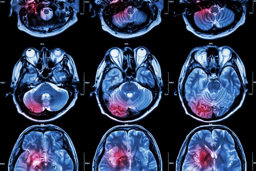
A useful brain imaging technique uses functional magnetic resonance imaging to analyse metabolic changes such as blood oxygenation.
Types Of Brain Imaging Techniques
Advancements in brain imaging using non-invasive technologies such as functional magnetic resonance imaging (fMRI), magnetoencephalography (MEG), and electroencephalography (EEG) have allowed neuroscientists to obtain a greater understanding of how the brain functions with its environment.
Understanding neuro-networks will assist in the development of treatments for neurodegenerative diseases such as Alzheimer’s and Parkinson’s. Various animal models have been developed to complement brain imaging to investigate the genetic nature of disease states, especially concentrating on neurophysiological processes.
A useful brain imaging technique uses functional magnetic resonance imaging (fMRI) by analysing metabolic changes such as blood oxygenation. The advantage of using fMRI is that it produces good spatial resolution. The disadvantage of fMRI is its poor time resolution of approximately six seconds and is therefore too slow for tracking single or clusters of neurons in real-time. This is because the change in blood flow takes several seconds to catch up with neural activity. However, fMRI has good spatial specificity in localised brain function compared to EEG, which uses electrical and magnetic signals derived from MEG.
Consequently, both EEG and MEG have excellent temporal resolution but very poor spatial precision. This is even though these imaging techniques can track neuron population activity within several hundreds of milliseconds. During these processes, neither imaging modalities can distinguish which set of neurons is being used.
To circumvent these issues, fMRI is sometimes used in conjunction with EEG to obtain paramount temporal and spatial precision.
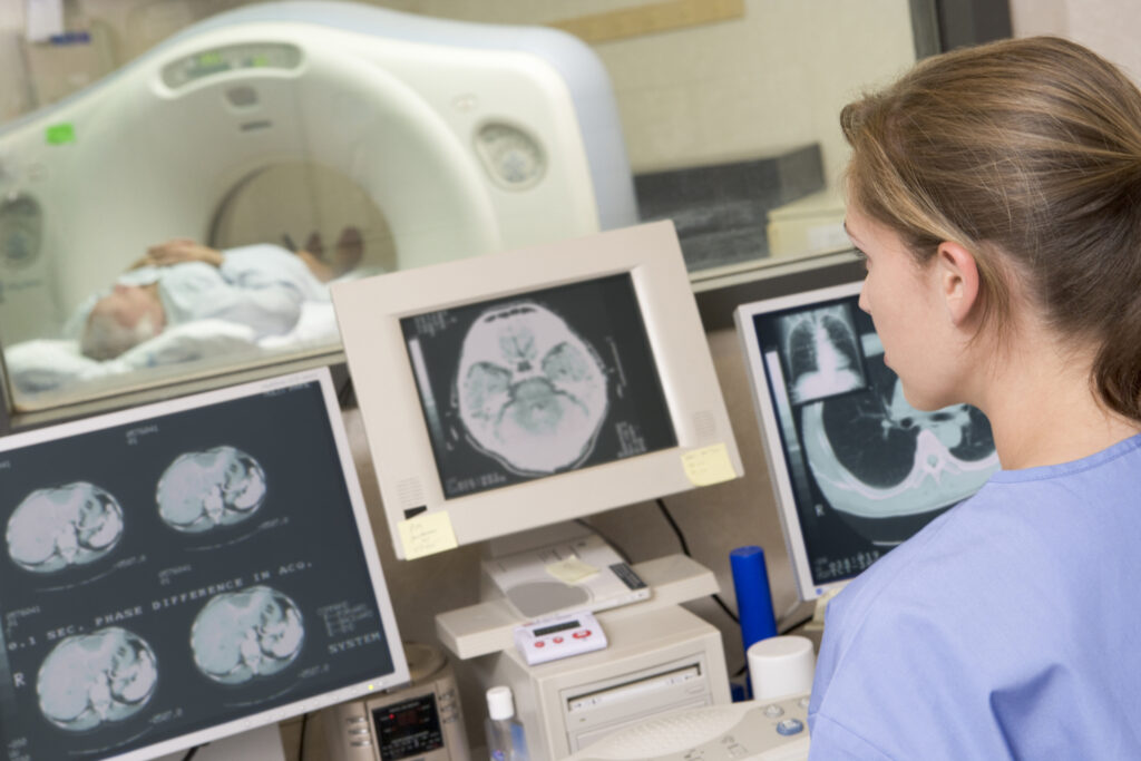
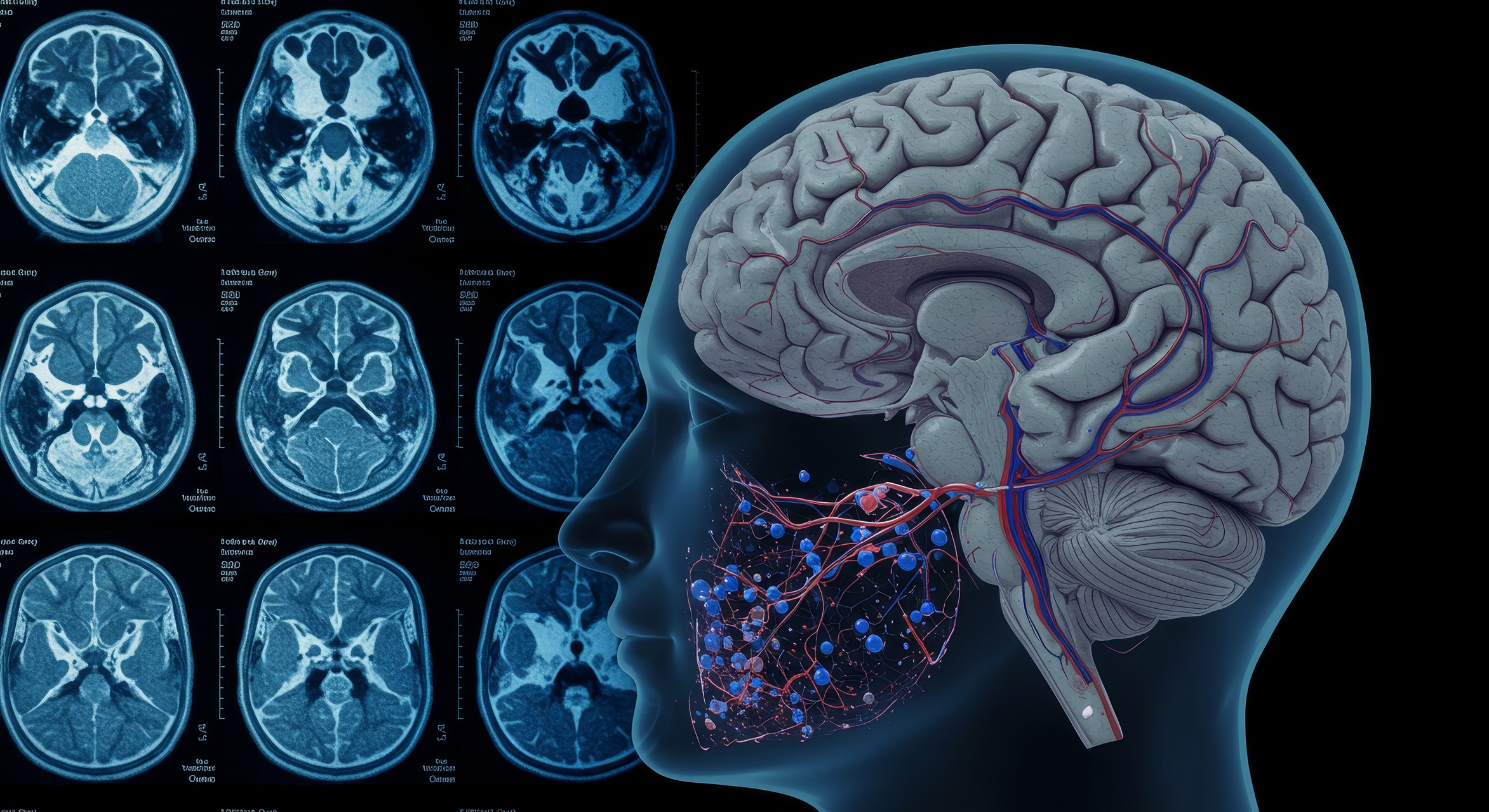
Brain imaging can make a contribution to the patient through personalised medicine in the treatment and management of neurological diseases by creating 3-D individual images in real-time. Both MRI and computed tomography (CT) hybrid scanners can be used to generate 3-D images of the brain at a specific moment.
These 3-D images of brain volume are made up of voxels.
The spatial resolution of the MRI scanner determines how small voxels can be measured during brain imaging of neural networks. Therefore, the stronger the magnetic field strength, the higher the spatial resolution and the better the resolution of brain structure.
Image-guided systems controlled by advanced brain navigation software will assist neurosurgeons in performing the operation on the patient in precise locations. These systems are based on the Talairach coordinates (x, y, and z).
Comparison Of Neuroimaging Modalities
S = Spatial Resolution; T = Temporal Resolution
Shriram R, Sundhararajan M, Daimiwal N. Brain connectivity analysis methods for better understanding of coupling. IJCSIS. 2012; 10(11).
Therefore, brain imaging assists neurosurgeons in removing disease-causing agents and preserving the function of the surrounding tissues. These imaging modalities include MRI and CT, which generate a 3-D brain map in real time and is essential to understanding functional measurements using EEG, MEG, and Functional MRI Brain Imaging, including deep brain recording and the ability to understand brain structure and function.
Extension to Types of Brain Imaging Techniques
Advancements in brain imaging using non-invasive technologies such as functional magnetic resonance imaging (fMRI), magnetoencephalography (MEG), and electroencephalography (EEG) have allowed neuroscientists to obtain a greater understanding of how the brain functions with its environment.
Understanding neuro-networks will assist in the development of treatments for neurodegenerative diseases such as Alzheimer’s and Parkinson’s. Various animal models have been developed to complement brain imaging to investigate the genetic nature of disease states, especially concentrating on neurophysiological processes.
A useful brain imaging technique uses functional magnetic resonance imaging (fMRI) by analysing metabolic changes such as blood oxygenation. The advantage of using fMRI is that it produces good spatial resolution. The disadvantage of fMRI is its poor time resolution of approximately six seconds and is therefore too slow for tracking single or clusters of neurons in real-time. This is because the change in blood flow takes several seconds to catch up with neural activity. However, fMRI has good spatial specificity in localised brain function compared to EEG, which uses electrical and magnetic signals derived from MEG.
Consequently, both EEG and MEG have excellent temporal resolution but very poor spatial precision. This is even though these imaging techniques can track neuron population activity within several hundreds of milliseconds. During these processes, neither imaging modalities can distinguish which set of neurons is being used.
To circumvent these issues, fMRI is sometimes used in conjunction with EEG to obtain paramount temporal and spatial precision.
Brain imaging can help the patient through personalized medicine in treating and managing neurological diseases by creating 3-D individual images in real time. MRI and computed tomography (CT) hybrid scanners can generate 3-D images of the brain at a specific moment.
These 3-D images of brain volume are made up of voxels.
The spatial resolution of the MRI scanner determines how small voxels can be measured during brain imaging of neural networks. Therefore, the stronger the magnetic field strength, the higher the spatial resolution and the better the resolution of brain structure.
Image-guided systems controlled by advanced brain navigation software will assist neurosurgeons in performing the operation on the patient in precise locations. These systems are based on the Talairach coordinates (x, y, and z).
Advancements in Non-Invasive Brain Imaging Techniques
Further advancements in non-invasive brain imaging technologies continue to revolutionise our understanding of the brain. One such advancement is the development of diffusion tensor imaging (DTI), a type of MRI that enables the mapping of the diffusion process of molecules, mainly water, in biological tissues. DTI is particularly useful for visualising white matter tracts and assessing the integrity of neural pathways, which is crucial in studying brain disorders such as multiple sclerosis and traumatic brain injury.
Positron Emission Tomography (PET)
Another significant technique is positron emission tomography (PET), which allows for imaging metabolic processes in the brain by detecting gamma rays emitted by a radioactive tracer. PET is especially beneficial in the early diagnosis and monitoring of diseases like Alzheimer’s, as it can detect amyloid plaques and tau tangles, which are hallmarks of the disease. Combining PET with MRI (PET-MRI) provides both metabolic and structural information, offering a comprehensive view of the brain’s condition.
Functional Near-Infrared Spectroscopy (fNIRS)
Functional near-infrared spectroscopy (fNIRS) is an emerging technology that measures brain activity by detecting changes in blood oxygenation and volume. This technique is portable and less expensive than fMRI, making it accessible for a wider range of research and clinical settings. fNIRS is particularly useful in studying brain function in infants and young children, as it is more tolerant of movement than other imaging modalities.
Advances in Computational Techniques
Advances in computational techniques and machine learning algorithms have also enhanced brain imaging analysis. These methods enable the processing of vast amounts of imaging data to identify patterns and correlations that may not be apparent through traditional analysis. Machine learning can improve the accuracy of disease diagnosis, predict patient outcomes, and personalise treatment plans based on individual brain imaging profiles.
Conclusion
In conclusion, the continuous development and refinement of brain imaging techniques are providing deeper insights into the structure and function of the brain. These advancements improve our understanding of neurophysiological processes and pave the way for more effective diagnosis, treatment, and management of neurological diseases. The integration of various imaging modalities and computational advancements holds great promise for the future of neuroscience and personalised medicine.
Disclaimer
The information provided in this article is intended for general informational and educational purposes only. It is not a substitute for professional medical advice, diagnosis, or treatment. While efforts have been made to ensure the accuracy of the content at the time of publication, Open Medscience makes no guarantees regarding the completeness, reliability, or accuracy of the information presented.
Any medical or scientific information referenced should be verified with qualified healthcare or research professionals. Readers should not disregard professional medical advice or delay seeking it because of something they have read on this website.
Open Medscience does not endorse or recommend any specific tests, procedures, opinions, or other information mentioned in this article. The views expressed are those of the original authors and do not necessarily reflect the views of Open Medscience.
Use of this article and reliance on any information provided is solely at your own risk.