Introduction
Modern Medical Imaging and Radiation Therapy (2024), written by Dr Sean L Kitson and published by Open MedScience, presents a wide-ranging exploration of diagnostic imaging and oncological treatment in contemporary healthcare. The book positions medical imaging and radiation therapy as interconnected disciplines that together define modern precision medicine.
Structured as an expansive compendium rather than a narrowly focused textbook, the volume integrates imaging physics, radiation biology, artificial intelligence, cybersecurity, robotics, and molecular theranostics into a unified examination of how technology continues to reshape patient care.
Foundations of Medical Imaging
The book begins by outlining the historical and scientific development of medical imaging. From the discovery of X-rays in 1895 to today’s advanced multimodal systems, this timeline traces the technological progression that enables clinicians to visualise internal anatomy without invasive intervention.
Core imaging modalities are explained clearly and methodically. X-ray and computed tomography (CT) are presented as foundational tools in trauma care, skeletal assessment, and oncological staging. The principle of differential absorption is described in accessible terms, demonstrating how tissue density differences produce diagnostic contrast.
Magnetic resonance imaging (MRI) is considered a non-ionising modality particularly suited to soft-tissue evaluation. Its application in neurological disorders, musculoskeletal injury, and cardiovascular disease highlights its diagnostic versatility. Ultrasound is described as a real-time, radiation-free technique widely used in obstetrics, cardiology, and abdominal diagnostics.
Nuclear medicine, including PET and SPECT, expands imaging beyond structural representation to physiological and molecular processes. Radiotracers are shown to reveal metabolic activity, enabling early detection and more precise disease characterisation.
Advances in CT and MRI Technology
Significant attention is devoted to technological refinement in CT and MRI systems. Developments such as multi-slice CT scanners, iterative reconstruction algorithms, low-dose protocols, and photon-counting CT demonstrate how improvements in image clarity have been accompanied by greater emphasis on radiation safety.
MRI coverage extends into functional and quantitative imaging, including diffusion-weighted imaging and functional MRI techniques. These developments illustrate how MRI now contributes not only to anatomical diagnosis but also to understanding tissue function and disease mechanisms.
Hybrid imaging platforms combining PET with CT or MRI are highlighted as transformative advances. By merging anatomical detail with metabolic data, these systems enhance staging accuracy and treatment planning, particularly in oncology and cardiology.
Nuclear Medicine and Theranostics
A defining strength of the book lies in its detailed exploration of nuclear medicine and molecular imaging. PET and SPECT are presented as modalities capable of mapping biochemical pathways and receptor expression, moving imaging into the domain of cellular biology.
Theranostics, which links molecular imaging with targeted radionuclide therapy, receives particular focus. By using similar molecular targets for both diagnosis and treatment, theranostics embodies personalised oncology. Targeted alpha and beta therapies are discussed as examples of precision treatment strategies guided by imaging science.
This section reflects a broader shift in medicine toward biologically informed, individualised interventions.
Radiation Therapy: Precision and Innovation
Radiation therapy is explored as the therapeutic counterpart to diagnostic imaging. High-energy particles or waves are described as mechanisms for disrupting cancer cell DNA and inhibiting tumour growth. External beam radiotherapy, brachytherapy, stereotactic radiosurgery, and proton therapy are examined within both biological and technological contexts.
Advanced techniques such as intensity-modulated radiation therapy (IMRT) and image-guided radiation therapy (IGRT) illustrate how radiation delivery has become increasingly precise. Imaging plays a critical role in mapping tumour geometry, verifying patient positioning, and refining treatment accuracy.
Proton beam therapy is presented as an example of highly controlled dose distribution, particularly beneficial in anatomically sensitive cases. Throughout, the integration of imaging data into radiotherapy planning underscores the personalised nature of modern cancer treatment.
Artificial Intelligence and Big Data
A central theme throughout the book is the integration of artificial intelligence and big data into imaging and oncology workflows. Deep learning algorithms, convolutional neural networks, and automated reconstruction techniques are described as tools that enhance image quality and support diagnostic interpretation.
Radiomics is presented as an emerging field that extracts quantitative features from medical images to predict prognosis and treatment response. Imaging is reframed not only as visual analysis but as data-rich information capable of supporting predictive modelling.
The discussion extends to structured and unstructured healthcare data environments, positioning imaging within a broader digital ecosystem. Artificial intelligence is portrayed as an enhancement to clinical expertise, improving efficiency and analytical depth rather than replacing professional judgement.
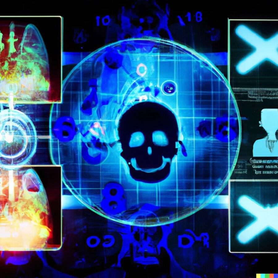
Cybersecurity and Digital Infrastructure
An especially forward-thinking aspect of the book is its attention to cybersecurity. As imaging systems, archives, and radiation therapy devices become increasingly networked, vulnerability to cyber threats grows.
The book addresses risks such as ransomware attacks, data breaches, and potential device compromise. Emphasis is placed on encryption, system monitoring, and staff education to protect patient data and treatment integrity. By including digital security within its scope, the work acknowledges that modern patient safety extends beyond biological risk to encompass technological resilience.
Clinical Applications Across Specialties
Imaging and radiation therapy are contextualised across a wide range of medical specialties. Dedicated discussions cover liver disease, cardiac imaging, skeletal disorders, breast cancer screening, neurological pathology, prostate cancer management, and neurodegenerative disease.
Low-dose CT for lung cancer screening, PET imaging in dementia research, and advanced radiotherapy techniques for metastatic disease demonstrate how technological advances translate into practical clinical benefit. The specialty-based approach reinforces the versatility and impact of modern imaging systems.
Emerging Technologies and Future Directions
Beyond established modalities, the book explores robotics, laser medicine, deep learning reconstruction, dark-field CT, phase-contrast imaging, and photon-counting detector systems. These chapters highlight emerging technologies that may redefine diagnostic resolution and therapeutic precision.
Robotic and image-guided interventions demonstrate the convergence of imaging with minimally invasive treatment. Advanced detector technologies promise improved contrast with reduced radiation exposure. The inclusion of such innovations reflects a field that continues to evolve rapidly.
Conclusion
Modern Medical Imaging and Radiation Therapy offers a comprehensive and forward-looking examination of diagnostic imaging and cancer treatment in the mid-2020s. By integrating foundational science, clinical application, digital transformation, and emerging molecular strategies, the book reflects the interdisciplinary nature of modern healthcare.
It presents medical imaging and radiation therapy not as static technologies, but as dynamic systems central to precision medicine, technological innovation, and improved patient outcomes.
home »