In medical diagnostics, the ability to see beneath the surface of tissues without invasive procedures has always been a coveted goal. Optical Coherence Tomography (OCT) represents a significant leap towards this goal, providing clinicians and researchers with detailed images of tissue microstructures in real time. First introduced in the early 1990s, OCT has undergone remarkable technological advancements, expanding its application far beyond ophthalmology into various medical fields. This article explores the fundamentals of OCT, its evolution, and its multifaceted applications.
Fundamentals of Optical Coherence Tomography
Optical Coherence Tomography is an imaging modality akin to ultrasound, but instead of sound waves, it employs light waves to capture images. The principle behind OCT is low-coherence interferometry, where light is split into a reference beam and a sample beam. The sample beam reflects off the tissue and combines with the reference beam, creating an interference pattern that is used to construct detailed images.
Working Principle
OCT utilises a broadband light source to penetrate tissues, typically in the near-infrared spectrum. The light source emits a low-coherence beam that is split into two paths. One path is directed towards the tissue, while the other is reflected off a reference mirror. The reflected light from both paths is recombined to produce an interference pattern, which is analysed to generate a high-resolution, cross-sectional image of the tissue.
Types of OCT
- Time-Domain OCT (TD-OCT): The original form of OCT, TD-OCT, involves mechanically moving the reference mirror to acquire depth information, which is relatively slow.
- Spectral-Domain OCT (SD-OCT): An advancement over TD-OCT, SD-OCT uses a stationary reference mirror and a spectrometer to capture the interference pattern, resulting in faster image acquisition and higher resolution.
- Swept-Source OCT (SS-OCT): This type employs a tunable laser that sweeps through a range of wavelengths, allowing deeper tissue penetration and providing even faster imaging than SD-OCT.
Technological Advancements
Significant improvements in image resolution, acquisition speed, and tissue penetration depth have marked the evolution of OCT technology, broadening the scope of its applications in medicine.
Modern OCT systems can achieve axial resolutions of 1-15 micrometres and lateral resolutions of 10-20 micrometres. The transition from TD-OCT to SD-OCT and SS-OCT has substantially increased the imaging speed, enabling real-time imaging and reducing motion artefacts.
Functional Extensions
- Optical Coherence Angiography (OCTA): A non-invasive technique for visualising blood flow within tissues, particularly useful in detecting retinal vascular diseases.
- Polarisation-sensitive OCT (PS-OCT) enhances tissue characterisation by detecting changes in the polarisation state of light, aiding in differentiating between various tissue types.
- Doppler OCT: Measures blood flow velocity by analysing frequency shifts in the reflected light, which is beneficial in assessing vascular conditions.
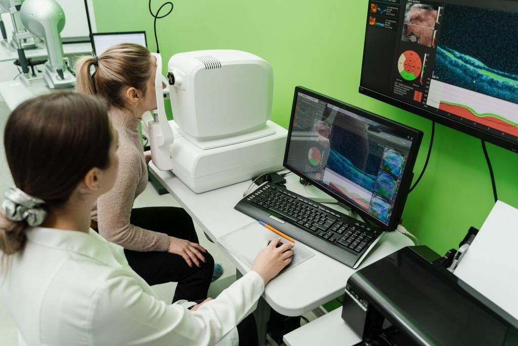
Applications in Ophthalmology
OCT has become an indispensable tool in ophthalmology. It provides detailed images of the eye’s retina, optic nerve, and anterior segment, aiding in the diagnosis and management of various eye conditions.
Retinal Diseases
OCT is particularly valuable in diagnosing and monitoring retinal diseases such as age-related macular degeneration (AMD), diabetic retinopathy, and macular oedema. It allows for the early detection of these conditions, enabling timely intervention and better patient outcomes.
Glaucoma
In glaucoma management, OCT is used to measure the thickness of the retinal nerve fibre layer (RNFL) and the optic nerve head, helping to detect glaucomatous damage at an early stage.
Anterior Segment Imaging
OCT provides detailed images of the cornea, iris, and anterior chamber, assisting in the evaluation of corneal diseases, angle-closure glaucoma, and pre- and post-operative assessment of refractive surgery.
Beyond Ophthalmology: Expanding Horizons
While OCT’s role in ophthalmology is well-established, its applications extend to various other medical fields, offering valuable insights into tissue microstructures and pathological changes.
Cardiology
In cardiology, OCT is used to image coronary arteries, providing high-resolution images of arterial walls and plaques. This aids in the assessment of atherosclerosis and guides interventions such as angioplasty and stent placement.
Dermatology
OCT helps in diagnosing skin conditions by providing detailed images of the skin’s microstructure. It is particularly useful in identifying basal cell carcinoma, squamous cell carcinoma, and other skin lesions without the need for a biopsy.
Oncology
In oncology, OCT is employed to image tissues and detect early signs of cancer. It aids in the evaluation of tumour margins during surgery, ensuring complete removal of cancerous tissues while preserving healthy tissue.
Gastroenterology
OCT is also utilised in gastroenterology to image the gastrointestinal tract, aiding in the detection and assessment of conditions such as Barrett’s oesophagus and colorectal cancer.
Future Prospects and Innovations
The future of OCT is promising, with ongoing research and technological innovations poised to further expand its capabilities and applications.
Integration with Artificial Intelligence
The integration of OCT with artificial intelligence (AI) holds the potential to enhance diagnostic accuracy and efficiency. AI algorithms can assist in the automated analysis of OCT images, identifying subtle changes and patterns that may be overlooked by the human eye.
Multimodal Imaging
Combining OCT with other imaging modalities, such as fluorescence microscopy or photoacoustic imaging, can provide complementary information, offering a more comprehensive view of tissue structures and functions.
Portable and Miniaturised OCT Devices
The development of portable and miniaturised OCT devices could make this technology more accessible, particularly in remote and resource-limited settings. This would enable widespread screening and monitoring of various conditions, improving healthcare delivery.
Conclusion
Optical Coherence Tomography has undeniably transformed the landscape of medical diagnostics, offering unparalleled insights into the microstructures of biological tissues. From its origins in ophthalmology to its expanding applications in cardiology, dermatology, oncology, and beyond, OCT continues to be a vital tool in the early detection and management of numerous conditions. With ongoing advancements and innovations, the future of OCT promises even greater capabilities and broader accessibility, solidifying its role as a cornerstone of modern medical imaging. As technology progresses, OCT will likely uncover even more of the intricate details of human health, contributing to improved patient outcomes and advancing the field of medicine.
Disclaimer
The information presented in this article is intended for educational and informational purposes only. While every effort has been made to ensure accuracy, the content does not constitute medical advice, diagnosis, or treatment and should not be relied upon as such. Readers are advised to consult qualified healthcare professionals for any medical concerns or conditions. The views expressed are those of the authors and do not necessarily reflect those of any affiliated institutions or organisations. Neither the authors nor the publisher assume any responsibility or liability for any loss, injury, or damage resulting from the use or misuse of the information provided.
home »