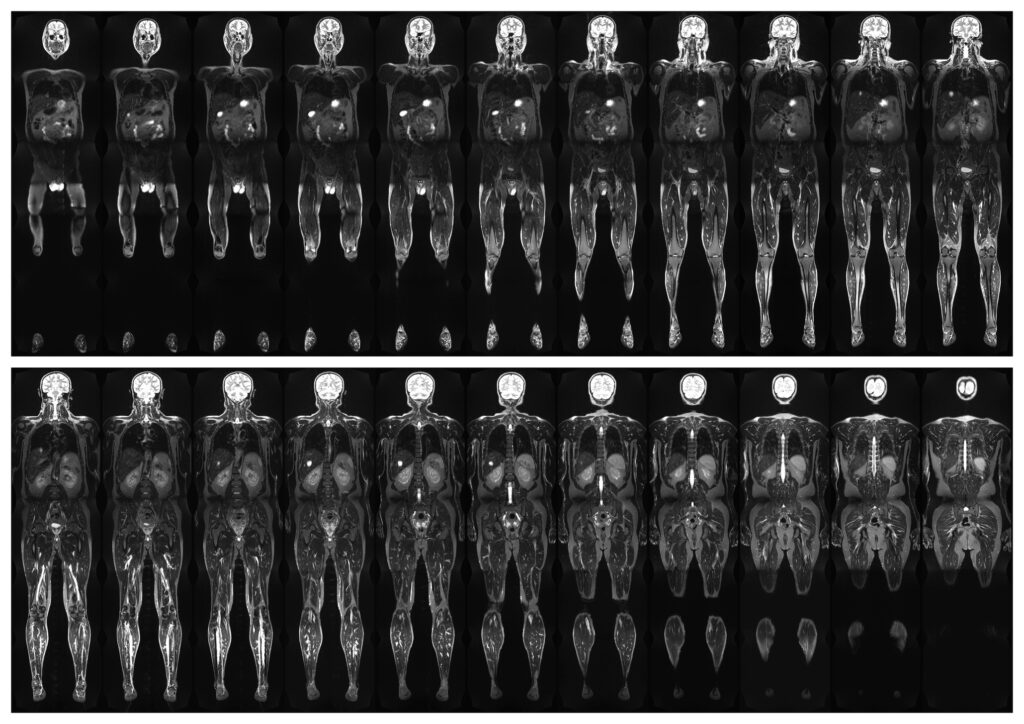
Quantitative Whole-Body MRI: A Transformative Approach to Systemic Imaging
 By
By
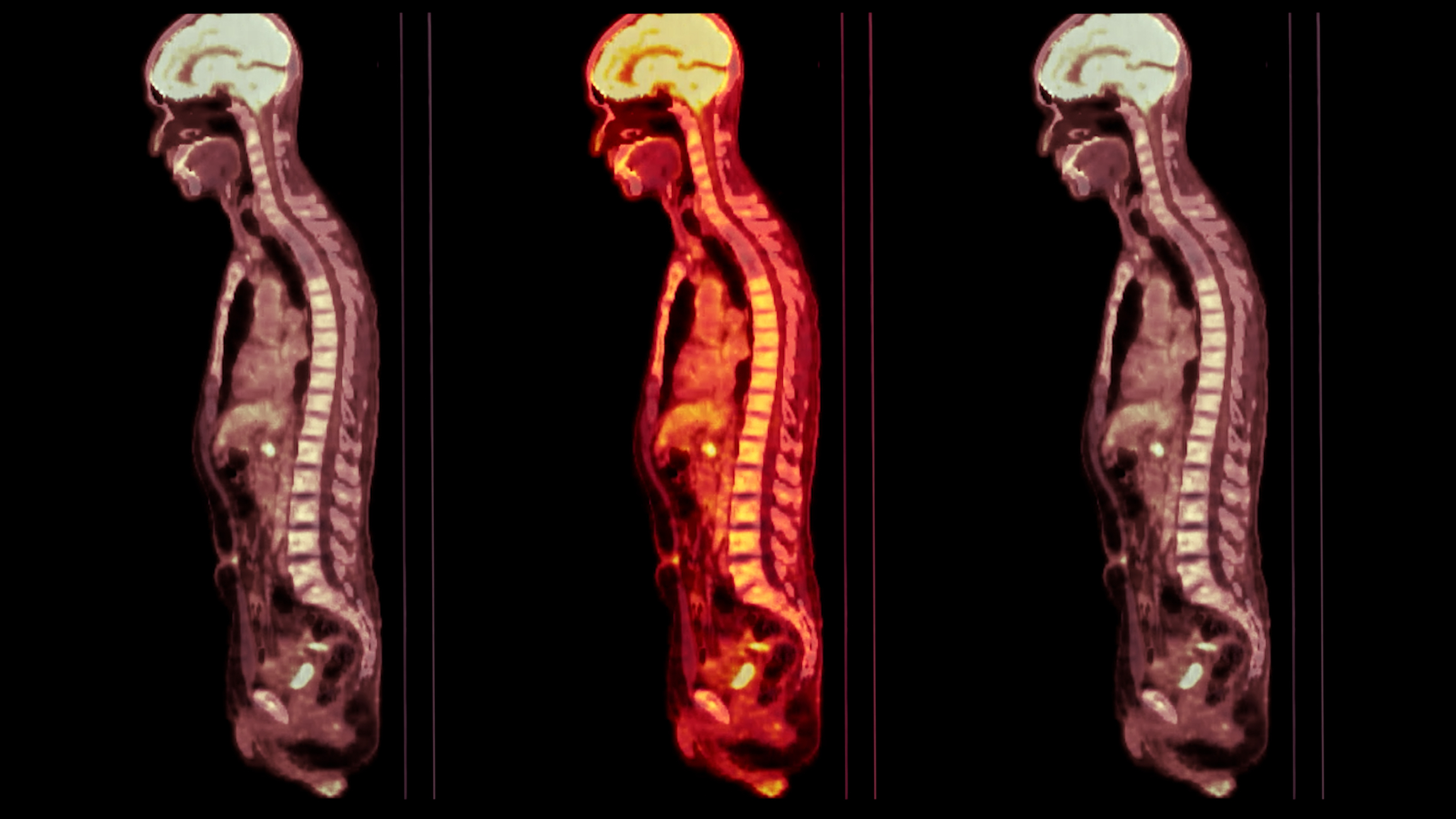
Understand the principles of Quantitative Whole-Body MRI and its applications in modern medicine for effective disease assessment.
Medical imaging topics encompass a broad spectrum of technologies that underpin modern diagnosis and treatment. Core modalities include X-ray, computed tomography (CT), magnetic resonance imaging (MRI), ultrasound and nuclear medicine. X-ray imaging remains essential for assessing fractures and chest pathology, while CT provides detailed cross-sectional and three-dimensional visualisation for trauma, oncology and complex anatomical evaluation. MRI uses magnetic fields and radiofrequency signals to generate high-contrast images of soft tissues, supporting neurological, musculoskeletal and oncological assessment without ionising radiation. Ultrasound provides real-time imaging using sound waves and is widely used in obstetrics, vascular medicine, and abdominal diagnostics. Nuclear medicine employs radiotracers and gamma or PET imaging to reveal physiological and metabolic processes, enabling functional assessment of cancer, cardiac disease and neurological disorders.
home »
By
Understand the principles of Quantitative Whole-Body MRI and its applications in modern medicine for effective disease assessment.
 By
By
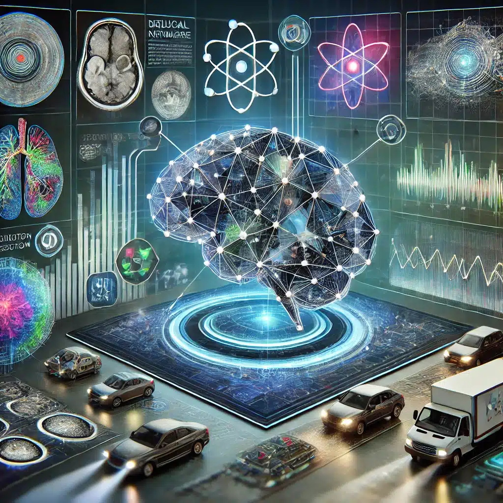
Artificial Intelligence and Machine Learning are revolutionising image analysis, enhancing precision, automating decisions, and driving innovation across industries such as healthcare, security, and autonomous systems.
 By
By
Diagnostic imaging in motor neurone disease (MND) is crucial for early detection, disease monitoring, and differentiating from other conditions.
 By
By
CAR T-cell therapy revolutionises cancer care, bringing hope where traditional treatments have been insufficient.
 By
By
SPECT imaging, by combining functional and anatomical insights, significantly enhances diagnostic accuracy in various medical fields.
 By
By
PET imaging dramatically enhances early disease detection, significantly improving patient outcomes in various medical fields.
 By
By
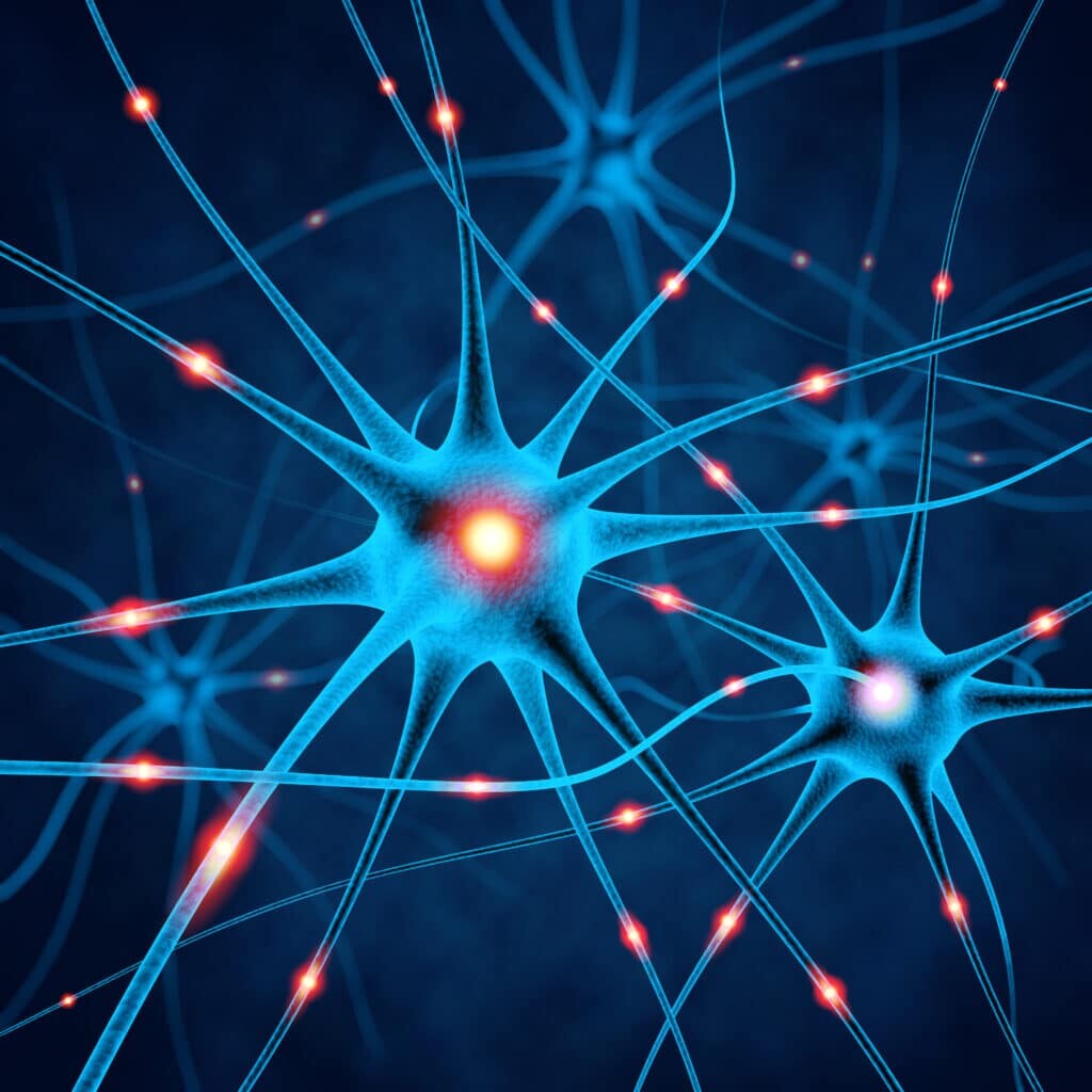
Neurological diagnostics have been transformed by advanced imaging techniques, enhancing accuracy in identifying brain disorders.
 By
By
Radiopharmaceuticals, combining radioactive and pharmaceutical elements, enable precise diagnosis and treatment in nuclear medicine.
 By
By
Theranostics merges diagnostics and therapy, revolutionising personalised medicine with genetic profiling and targeted treatment strategies.
 By
By
Medical imaging crucially enhances oncology, aiding early cancer detection and effective treatment planning. Image for illustration only. People depicted are models.
 By
By
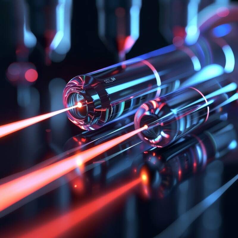
Lasers in medicine offer revolutionary precision and versatility, significantly advancing healthcare through diverse, minimally invasive applications.
 By
By
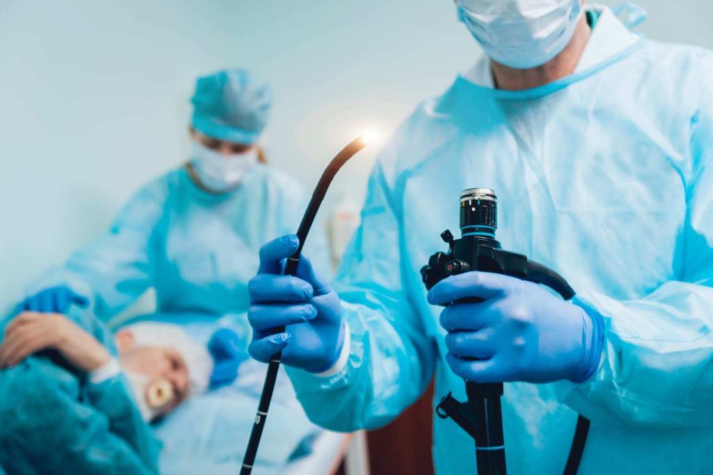
Fibre optics in endoscopy revolutionise medical procedures, enhancing diagnostics and surgical precision. Image for illustration only. People depicted are models.
 By
By
In Brazil, the growing radiopharmaceutical market, driven by chronic disease and advanced imaging technologies, is becoming increasingly significant in healthcare.
 By
By
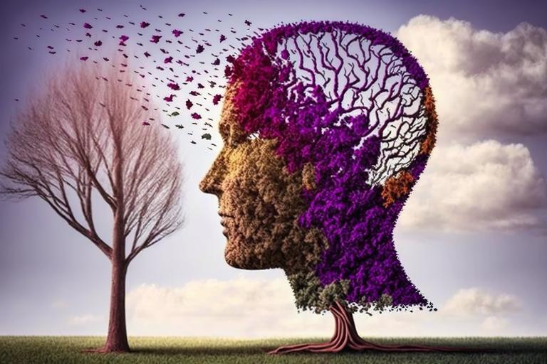
Alzheimer’s disease stems from genetic mutations and lifestyle factors, leading to brain plaque accumulation and dementia.
 By
By
Alzheimer’s disease, an age-related neurodegenerative disorder, impacts brain structure and function, requiring advanced imaging techniques for improved understanding and diagnosis.
 By
By
Medical laser milestones encompass pivotal discoveries, advancing treatment efficacy and expanding therapeutic applications.