
The Science Behind Quantum Wellness Technology
 By
By
Discover how quantum wellness technology enhances human vitality by utilizing principles from quantum physics and bioenergetics.
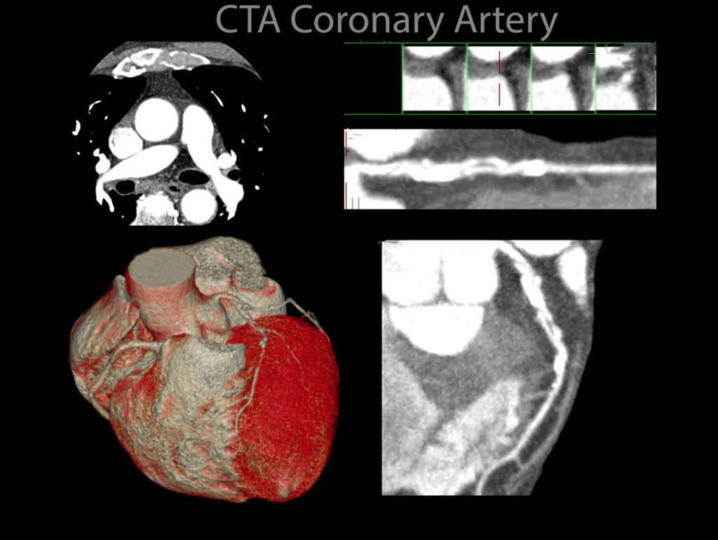
Cardiac electrophysiology imaging is a crucial tool in the study and diagnosis of heart rhythm disorders. It provides detailed insights into the electrical activity of the heart, helping clinicians diagnose arrhythmias, guide treatment strategies, and assess the effectiveness of interventions such as catheter ablation or implantable devices. Advances in imaging techniques have significantly improved the precision and accuracy of cardiac electrophysiology studies, allowing for better patient outcomes.
Fundamentals of Cardiac Electrophysiology
The heart’s electrical system controls the rhythm and coordination of contractions, ensuring efficient blood circulation. Electrical impulses originate from the sinoatrial (SA) node, travel through the atria, and pass through the atrioventricular (AV) node before reaching the ventricles. Disruptions in this electrical conduction can result in arrhythmias, which may be benign or life-threatening, depending on the nature of the disturbance.
Electrophysiological studies (EPS) are performed to evaluate abnormal electrical pathways. Traditionally, EPS involve invasive catheter-based recordings; however, non-invasive imaging techniques have gained traction, offering complementary or alternative approaches.
Imaging Techniques in Cardiac Electrophysiology
Several imaging modalities contribute to cardiac electrophysiology, each providing unique advantages:
Clinical Applications
Cardiac electrophysiology imaging plays a vital role in diagnosing atrial fibrillation, ventricular tachycardia, and other complex arrhythmias. It guides interventions such as catheter ablation, assists in risk stratification for sudden cardiac death, and evaluates the placement of implantable cardioverter defibrillators (ICDs) and cardiac resynchronisation therapy (CRT) devices.
As imaging technologies continue to evolve, their integration with artificial intelligence and advanced computational modelling holds promise for further refining diagnosis and treatment planning in cardiac electrophysiology.
home » Cardiac Electrophysiology Imaging
Discover how quantum wellness technology enhances human vitality by utilizing principles from quantum physics and bioenergetics.
Understand how AI-derived FFR-CT in the NHS is transforming the diagnosis of coronary artery disease and improving health equity.