
How Creative Hobbies Can Support Mental Wellbeing in Adults
 By
By
Discover the role of Creative Mental Wellbeing in promoting psychological health through engaging hobbies like painting and gardening.
By
Discover the role of Creative Mental Wellbeing in promoting psychological health through engaging hobbies like painting and gardening.
 By
By
Understand the role of Hair Growth Nutrition in combating hair loss. Discover essential nutrients for strong, healthy hair.
 By
By
Learn about alcohol use disorder treatment and the importance of evidence-based care in achieving lasting recovery.
 By
By
Find out how Complex Diagnosis Support can aid families navigating the healthcare and legal systems for medical conditions.
 By
By
Discover the best footwear for peripheral neuropathy to prevent injuries and enhance comfort while addressing sensory impairments.
 By
By
Discover the shift to evidence-based medicine in vein care, leading to personalized treatments and improved patient outcomes.
 By
By
Discover the impact of artificial intelligence in vascular diagnostics. Learn how machine learning improves blood flow analysis and diagnoses.
 By
By
Discover the benefits of EPA and DHA omega-3 fatty acids and how they contribute to overall well-being and health.
 By
By
Uncover the role of Omega-3 in maintaining brain function, memory, and concentration as part of a healthy diet.
 By
By
Gain insights into the development of ambulatory surgery centres and the overlapping phases that determine their successful opening.
 By
By
Understand the radiopharmaceutical supply chain and how it ensures effective delivery of essential medical treatments.
 By
By
Discover the emotional and financial toll of a serious injury. Understand its lasting impact on everyday life. Image for illustration only. People depicted are models.
 By
By
Recognizing Traumatic Brain Injury is vital. Learn about symptoms that may emerge days after a road accident.
 By
By
Discover how multimodal artificial intelligence transforms patient care by integrating various clinical data sources seamlessly.
 By
By
Understand the challenges of Clinical Imaging in identifying microscopic abnormalities that standard scans may miss.
 By
By
Learn about Oculoplastic Surgery and how it addresses eyelid concerns, improving both vision and facial aesthetics effectively. Image for illustration only. Person depicted is a model.
 By
By
Explore the vital role of medical documentation after injury. Proper records can affect treatment and future legal cases. Image for illustration only. People depicted are models.
 By
By
Learn everything about inguinal hernia surgery recovery, including tips on exercise, lifting, and returning to daily routines.
 By
By
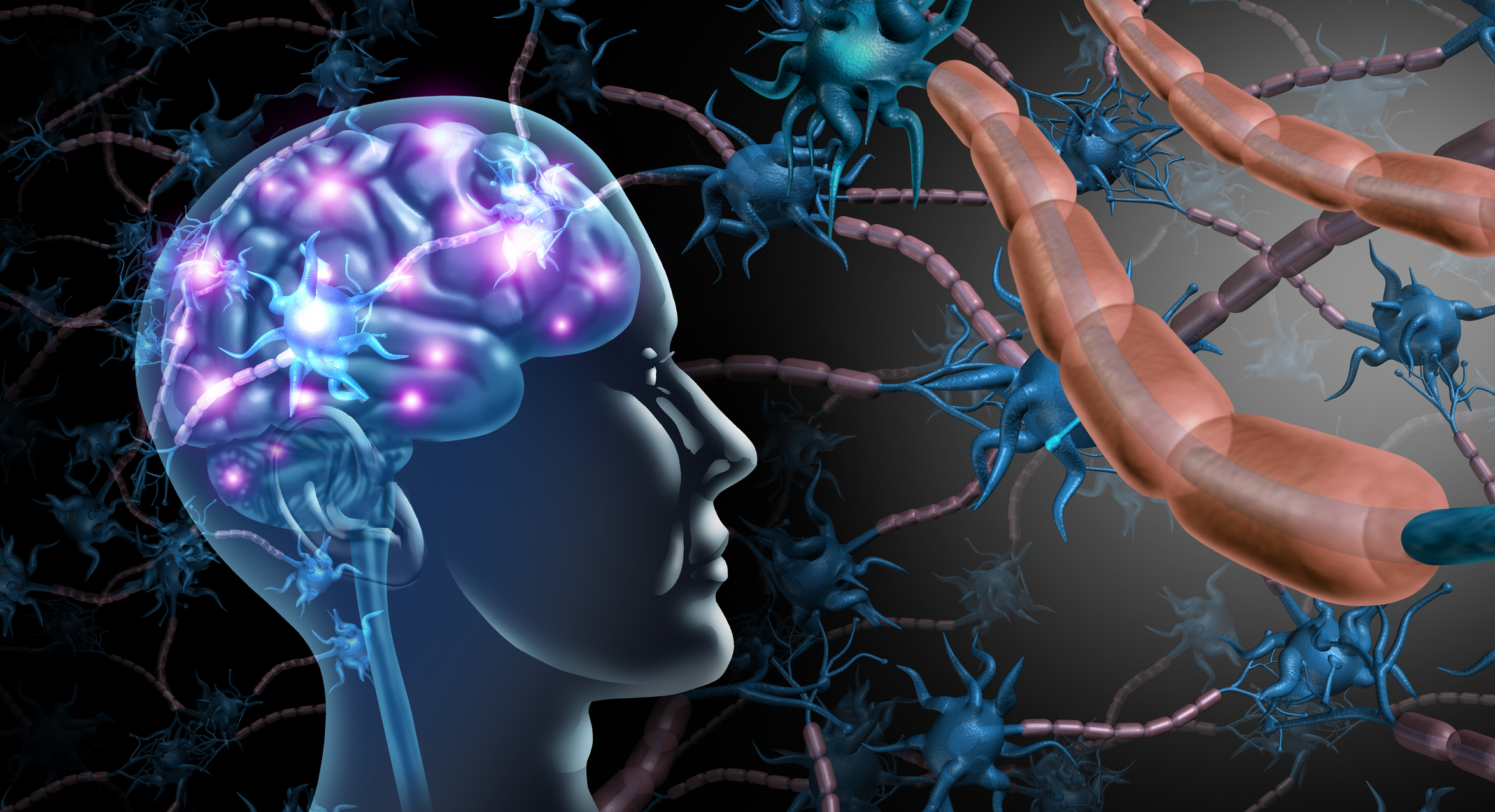
Uncover the breakthroughs in neuroscience research that are changing our perspective on brain activity and human behaviour.
 By
By
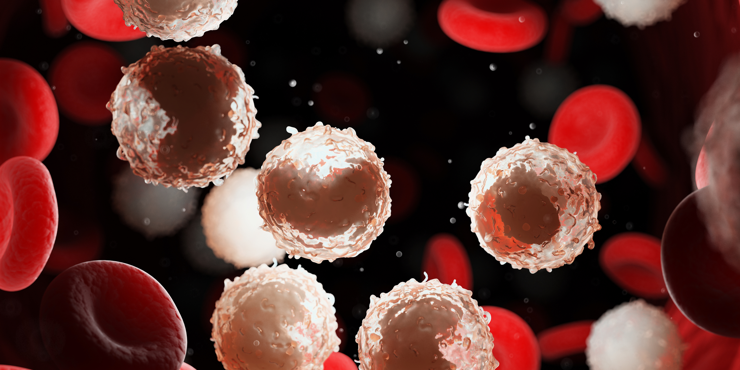
Explore the key factors in CLL treatment decisions and when to begin therapy for chronic lymphocytic leukaemia.
 By
By
Understand the role of radiolabelled peptides and molecular imaging in enhancing the precision of disease monitoring.
 By
By
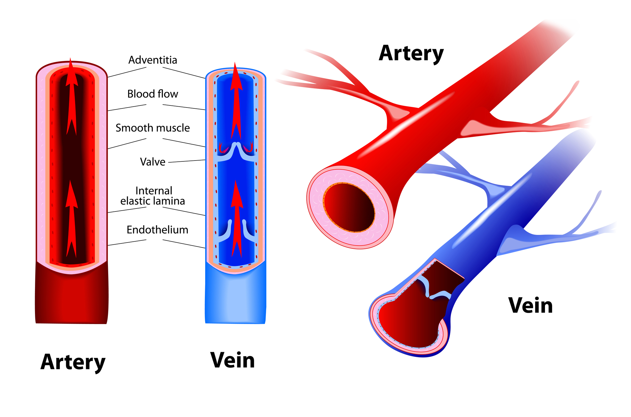
Explore chronic venous insufficiency symptoms that may be silently affecting your health. Learn to identify early signs.
 By
By
Discover how to locate primary care doctors near me efficiently, ensuring quality care and timely appointments for your needs. Image for illustration only. People depicted are models.
 By
By
Discover how custom psychiatry EMRs enhance behavioral health care documentation and improve clinician focus on patients. Image for illustration only. People depicted are models.
 By
By
Discover the potential of Retatrutide weight loss, a new contender in obesity drug therapy targeting multiple hormone receptors.
 By
By
Fungal infections require prompt treatment with appropriate antifungal therapy to reduce symptoms, prevent recurrence and support healthy skin.
 By
By
Learn how Mesothelioma medical imaging helps detect this rare cancer early and understand its progression effectively.
 By
By
Understand the importance of dietary supplements safety. Discover how science and regulation can guide your supplement choices.
 By
By
Find out everything you need to know about longevity clinics abroad and make informed choices for optimising your health.
 By
By
Learn about the role of online pharmacies in modern healthcare and their impact on medication management and accessibility.