How New MRI Contrast Agents Are Transforming Medical Imaging
 By
By
Uncover the impact of new MRI contrast agents on diagnostic imaging and their significance in modern medical technology.
Contrast agent dynamics modelling is a crucial aspect of medical imaging, particularly in techniques such as magnetic resonance imaging (MRI), computed tomography (CT), and positron emission tomography (PET). The primary aim is to understand how contrast agents behave within biological systems, enabling the accurate assessment of tissue perfusion, vascular integrity, and other physiological parameters.
Fundamentals of Contrast Agents
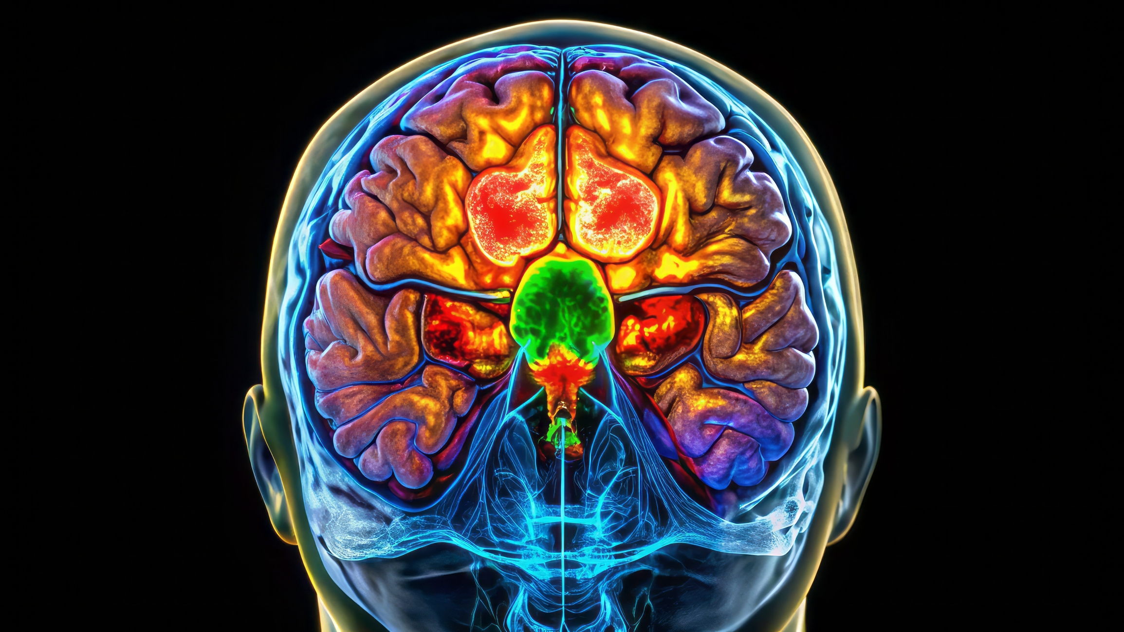
Contrast agents are substances introduced into the body to enhance imaging contrast. Their function is to alter signal intensity, either by increasing or decreasing the absorption, scattering, or emission of signals in response to the imaging modality used. In MRI, contrast agents such as gadolinium-based compounds influence relaxation times, whereas iodinated contrast agents in CT enhance X-ray attenuation. In PET, radiotracers such as fluorodeoxyglucose (FDG) are used to trace metabolic activity.
Mathematical Modelling of Contrast Agent Kinetics
Mathematical models describe how contrast agents move through the vascular and extravascular compartments. The two most commonly used models are the compartmental model and the distributed model.
The compartmental model simplifies tissue into well-mixed compartments where contrast agents enter, diffuse, and exit. The most basic form is the two-compartment model, which considers an intravascular and an extravascular-extracellular space (EES). The behaviour of the contrast agent is typically expressed using differential equations, with parameters such as permeability, flow rate, and volume fraction dictating the agent’s kinetics.
The distributed model offers a more detailed representation by incorporating spatial variations in contrast agent concentration. These models rely on partial differential equations (PDEs) and are often used in advanced simulations of contrast perfusion.
Applications in Medical Imaging
Contrast agent dynamics modelling is widely applied in diagnosing and monitoring diseases. In dynamic contrast-enhanced MRI (DCE-MRI), models quantify vascular permeability and extracellular volume fraction, aiding in tumour grading and treatment response assessment. Similarly, in CT perfusion imaging, contrast dynamics help identify ischaemic regions in stroke patients by measuring blood flow, blood volume, and mean transit time.
In PET imaging, kinetic modelling is employed to interpret tracer uptake and metabolism. This is particularly important in oncology, where the uptake of FDG can provide insights into tumour aggressiveness and response to therapy.
Challenges and Future Directions
Despite its significance, contrast agent dynamics modelling presents several challenges. The accuracy of models depends on high-quality data, which can be affected by motion artefacts, tracer dispersion, and physiological variability. Additionally, assumptions made in simpler models may not fully capture complex tissue heterogeneity.
Future developments focus on improving model accuracy through artificial intelligence and machine learning approaches. Advanced computational techniques can refine parameter estimation, reduce noise, and enhance real-time decision-making in clinical settings.
Contrast agent dynamics modelling remains a vital tool in medical imaging, offering deeper insights into physiological processes and contributing to more precise diagnoses and treatment planning.
home » Contrast Agent Dynamics Modelling
Uncover the impact of new MRI contrast agents on diagnostic imaging and their significance in modern medical technology.