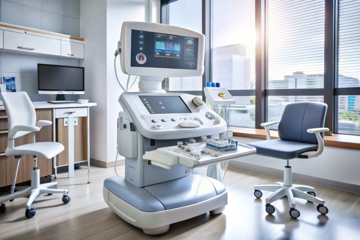
The Role of Ultrasound in Medical Imaging
Ultrasound provides non-invasive, real-time imaging, making it an essential diagnostic tool in modern medical practice.
Cystography is a specialised radiographic examination designed to evaluate the bladder’s anatomy and function. This diagnostic technique is essential for detecting and understanding various urological conditions, including bladder ruptures, vesicoureteral reflux, and bladder tumours.
The process involves introducing a contrast medium, typically a water-soluble iodine-based solution, into the bladder via a catheter. This contrast agent enhances the visibility of the bladder’s internal contours on X-ray images. There are two primary types of cystography: retrograde and voiding. Retrograde cystography fills the bladder with a contrast medium without the patient voiding, while voiding cystourethrography (VCUG) involves imaging during urination to assess both the bladder and the urethra.
Cystography is particularly valuable in paediatric medicine, where it helps diagnose congenital anomalies of the urinary tract, such as vesicoureteral reflux, which can lead to recurrent urinary tract infections and kidney damage if left untreated. In adults, this diagnostic tool is frequently utilised following traumatic injuries to the lower abdomen to confirm suspected bladder perforations.
The procedure begins with the patient lying on an X-ray table. After the bladder is filled with the contrast medium through the catheter, several X-ray images are taken from different angles to provide a comprehensive view of the bladder and surrounding structures. In VCUG, additional images are captured during and after the patient urinates to evaluate the function of the urinary tract and check for any abnormalities, such as the reverse flow of urine back towards the kidneys.
Although cystography is a highly informative diagnostic tool, it comes with potential risks, primarily related to the use of contrast materials. These risks include allergic reactions and infections. Hence, thorough sterilisation and adherence to procedural protocols are critical to minimise complications.
Moreover, advancements in imaging technology have led to the development of less invasive techniques, such as magnetic resonance (MR) cystography, which provides detailed images without radiation exposure. Nonetheless, traditional cystography remains indispensable in many clinical settings due to its accessibility and effectiveness in diagnosing complex urological disorders.
home » Cystography
Ultrasound provides non-invasive, real-time imaging, making it an essential diagnostic tool in modern medical practice.
