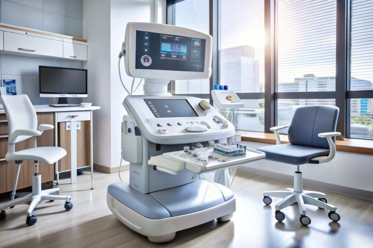
The Role of Ultrasound in Medical Imaging
Ultrasound provides non-invasive, real-time imaging, making it an essential diagnostic tool in modern medical practice.
Fetal Magnetic Resonance Imaging (MRI) is a revolutionary diagnostic tool that offers a deeper insight into prenatal development, surpassing the limitations of traditional ultrasound. This non-invasive technique provides high-resolution images of the fetus, placenta, and amniotic environment, making it an invaluable resource in assessing fetal health and diagnosing complex congenital anomalies.
Fetal MRI is typically recommended when ultrasound results are unclear or when there is a suspicion of abnormalities that require detailed assessment. It is particularly useful for evaluating brain development and detecting abnormalities in the central nervous system. This imaging technique is also beneficial in assessing congenital heart defects, chest anomalies, and gastrointestinal blockages. Unlike ultrasound, MRI does not use ionising radiation, making it safe for both the mother and fetus.
The procedure involves the use of strong magnetic fields and radio waves to generate detailed images of the body’s internal structures. During the scan, the expectant mother lies inside the MRI scanner, which is a large tube containing powerful magnets. The data collected are then transformed into detailed images by a computer. These images can be viewed in various planes, providing a comprehensive evaluation that ultrasound may not offer.
Despite its many benefits, fetal MRI is not without challenges. The primary limitation is the fetal movement, which can sometimes lead to blurred images. However, advances in technology have led to faster scanning techniques that minimise this issue and allow for clearer, more reliable imaging. Additionally, MRI is typically more expensive than ultrasound and may not be as readily available in all healthcare facilities.
Fetal MRI can provide critical information that influences the management of a pregnancy in cases where specific fetal conditions are suspected, such as ventriculomegaly, cleft lip, or spinal abnormalities. It assists healthcare professionals in planning necessary interventions, including surgeries that may be required immediately after birth. Furthermore, it helps in counselling parents about the expected outcomes and potential challenges in the child’s development.
In conclusion, fetal MRI represents a significant advancement in prenatal diagnostics. While it supplements rather than replaces ultrasound, its ability to provide clear, detailed images makes it an essential tool in the diagnosis of complex fetal conditions. As technology advances, the scope of fetal MRI is expected to expand, further enhancing its accuracy and utility in prenatal care. This progress holds the promise of improved outcomes for both mother and child, cementing its role in modern obstetrics.
home » Fetal MRI
Ultrasound provides non-invasive, real-time imaging, making it an essential diagnostic tool in modern medical practice.
