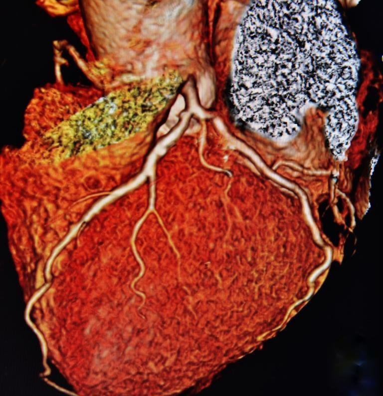
High Resolution CT
High-Resolution CT
High-resolution CT is an advanced imaging technique that provides detailed images of internal structures, primarily focusing on the lungs and airways. This method employs a special type of X-ray technology that captures extremely high-resolution images, enabling physicians to detect subtle abnormalities that may not be visible on standard CT scans.
High-resolution CT is particularly useful in diagnosing and monitoring diseases that affect the fine structure of the lungs, such as interstitial lung disease, fibrosis, and emphysema. By producing thin-slice images, typically 1-2 mm in thickness, HRCT offers an exquisite level of detail. These images can be viewed in both axial and coronal planes, giving a comprehensive view of the lung parenchyma and helping in the assessment of the airways, blood vessels, and the lung interstitium.
The procedure is usually quick, taking about 10-20 minutes to complete. It requires the patient to hold their breath for short intervals to minimize motion blur. Although HRCT involves exposure to radiation, the dose is generally lower compared to conventional CT scans, balancing the need for diagnostic accuracy with patient safety.
HRCT has become an indispensable tool in pulmonology, providing critical insights that guide treatment decisions. Its ability to pinpoint the location and nature of lung pathology supports a more accurate diagnosis, aids in determining the severity of lung diseases, and assists in monitoring disease progression or response to therapy. As imaging technology advances, the resolution and capabilities of HRCT continue to evolve, promising even greater contributions to medical diagnostics and patient care.
home » High-Resolution CT

