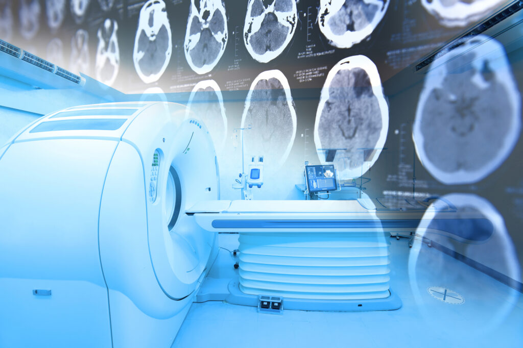
MRI Contrast Agents and Nutrition
Magnetic Resonance Imaging (MRI) is a widely used diagnostic tool that provides detailed images of internal organs, tissues, and structures. While conventional MRI scans rely on the natural differences in tissue composition, contrast agents enhance image clarity by altering the magnetic properties of tissues. The most commonly used MRI contrast agents are gadolinium-based, but concerns regarding their retention in the body have led to interest in alternative approaches, including the role of nutrition in optimising MRI scans.
MRI Contrast Agents
Gadolinium-based contrast agents (GBCAs) work by shortening the relaxation times of hydrogen protons in tissues, making them appear brighter on MRI scans. They are particularly useful for visualising blood vessels, detecting tumours, and assessing inflammatory or neurological conditions. However, some GBCAs may remain in the body for prolonged periods, particularly in individuals with kidney impairment, raising concerns about potential toxicity. To address this, macrocyclic GBCAs, which are more stable, have been developed to minimise retention.
In addition to gadolinium, iron oxide nanoparticles have been investigated as an alternative contrast agent. These particles, which consist of superparamagnetic iron oxides, are mainly used for liver imaging and lymph node assessments. They offer the advantage of being biodegradable and are eliminated through normal iron metabolism. Manganese-based contrast agents have also been explored, as manganese is a naturally occurring element with paramagnetic properties.
Nutrition and MRI Imaging
While contrast agents enhance imaging, dietary choices can also influence MRI scan quality. Certain nutrients impact tissue hydration, vascular integrity, and inflammation, all of which affect MRI results.
Hydration and MRI Clarity
Adequate hydration is essential for MRI accuracy, particularly for scans of the abdomen and pelvis. Water intake improves blood flow and reduces artefacts that may arise from dehydration. Conversely, excessive caffeine or alcohol consumption before an MRI scan may lead to dehydration, reducing the clarity of soft tissue imaging.
Iron and MRI Signals
Iron is a critical component of blood oxygenation, and its levels can influence the contrast in MRI scans. Low iron levels, seen in anaemia, can affect imaging of the brain and cardiovascular system, while excess iron deposits (as in haemochromatosis) may interfere with magnetic signals.
Antioxidants and Inflammation
Dietary antioxidants, such as vitamin C, vitamin E, and polyphenols from fruits and vegetables, can modulate inflammatory processes. Since inflammation affects tissue properties and contrast uptake, maintaining an anti-inflammatory diet may help standardise imaging conditions.
Fat Intake and Lipid Signals
High-fat meals before an MRI can influence imaging, particularly in scans assessing metabolic processes or fat distribution. For this reason, patients may be advised to avoid fatty foods before certain scans.
Although MRI contrast agents remain essential for detailed imaging, nutritional factors can support optimal imaging outcomes and may help reduce the reliance on exogenous contrast agents in some cases.
home » MRI Contrast Agents and Nutrition

 By
By