Medical Imaging Breakthroughs Driving the Future of Healthcare
 By
By
Stay updated on medical imaging breakthroughs that are transforming healthcare and improving patient outcomes in innovative ways.
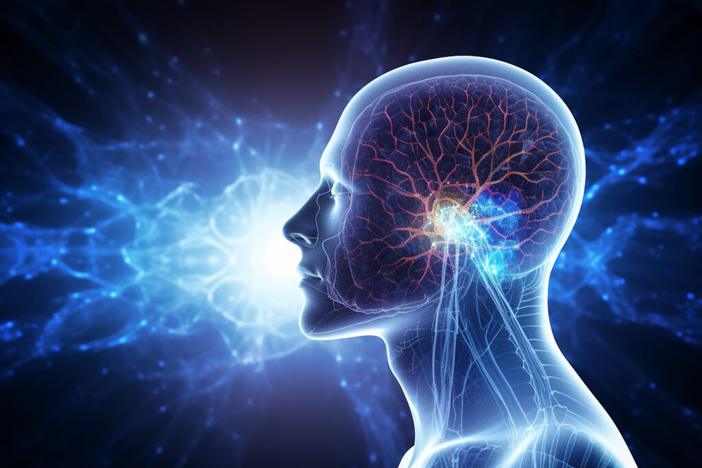
Positron Emission Tomography (PET) is a powerful imaging technique that provides functional insights into metabolic processes within the body. Unlike structural imaging methods such as CT or MRI, PET detects biochemical changes at the cellular level, making it particularly useful in fields such as oncology, neurology, and cardiology. By tracking radiolabelled molecules, PET enables the visualisation and quantification of metabolic pathways, offering valuable data for diagnosis, disease progression monitoring, and treatment evaluation.
Principles of PET Imaging
PET relies on the use of radiotracers—molecules labelled with positron-emitting radioisotopes such as fluorine-18 (18F), carbon-11 (11C), and oxygen-15 (15O). The most widely used radiotracer is fluorodeoxyglucose (18F^{18}F18F-FDG), a glucose analogue that accumulates in cells with high metabolic activity. When FDG is taken up by tissues, it undergoes phosphorylation but cannot proceed further in the glycolytic pathway, becoming trapped inside cells. This accumulation is detected by PET scanners, which measure positron annihilation events and construct a three-dimensional image of metabolic activity.
Metabolic Applications of PET
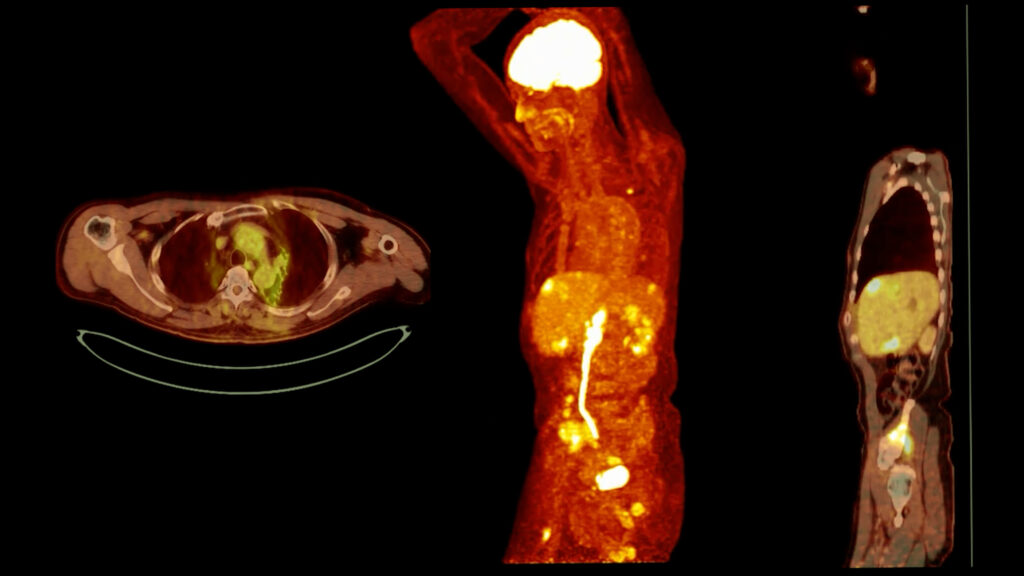
One of the most important applications of PET imaging is in oncology, where it exploits the altered metabolism of cancer cells. Malignant tumours often exhibit increased glucose uptake due to the Warburg effect—a phenomenon where cancer cells preferentially rely on glycolysis even in the presence of oxygen. FDG-PET can differentiate between benign and malignant lesions, assess tumour staging, and monitor treatment response. Reduced FDG uptake following chemotherapy or radiotherapy suggests effective treatment, while persistent or increased uptake may indicate residual disease.
In neurology, PET imaging is used to study cerebral metabolism, particularly in neurodegenerative disorders such as Alzheimer’s disease. Glucose metabolism in the brain is tightly regulated, and reductions in FDG uptake can indicate neuronal dysfunction. In Alzheimer’s, PET imaging typically reveals decreased metabolism in the temporoparietal and posterior cingulate cortices. Additionally, PET can be used to measure amyloid and tau deposition using specific radiotracers, aiding early diagnosis and disease characterisation.
In cardiology, PET is employed to assess myocardial metabolism and perfusion. The heart primarily utilises fatty acids for energy, but under conditions of stress or ischaemia, it shifts towards glucose metabolism. FDG-PET can identify viable but dysfunctional myocardium (hibernating myocardium) in patients with coronary artery disease, guiding revascularisation strategies. Other tracers, such as 11C-acetate and 13N-ammonia, provide additional insights into myocardial oxidative metabolism and perfusion.
Future Directions
Advances in PET imaging continue to refine our understanding of metabolism. Novel tracers are being developed to target specific metabolic pathways, such as amino acid transport, lipid metabolism, and hypoxia. Coupled with hybrid imaging modalities such as PET/MRI, these innovations promise to enhance diagnostic precision and expand PET’s role in personalised medicine.
PET imaging remains an indispensable tool for studying metabolism, offering unparalleled insights into disease mechanisms and treatment efficacy.
home »
By
Stay updated on medical imaging breakthroughs that are transforming healthcare and improving patient outcomes in innovative ways.
 By
By
Discover how health and wellbeing imaging insights are reshaping our understanding of metabolic health and personal wellness.
 By
By
Discover the advancements in PET imaging on-chip and its impact on preclinical research and radiotracer development.
 By
By
Explore total-body PET imaging and its advancements in sensitivity and clinical applications for better molecular insights.
 By
By
Understand the importance of PET imaging in healthcare, from cardiac assessments to patient journeys through nuclear medicine.
 By
By
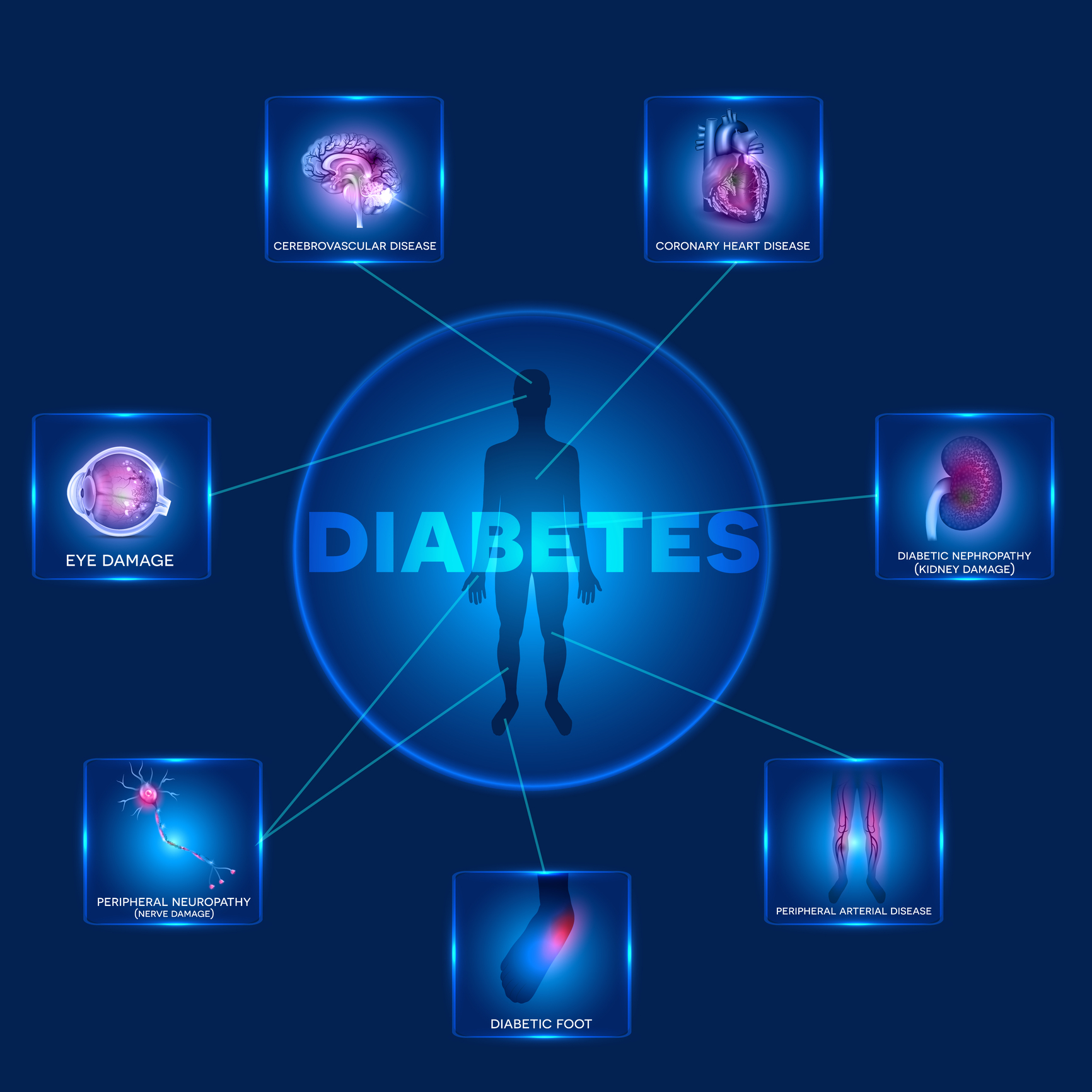
Different Types of Diabetes in medical imaging require tailored approaches for accurate diagnosis and effective complication monitoring.