A Short History of Magnetic Resonance Imaging (MRI)
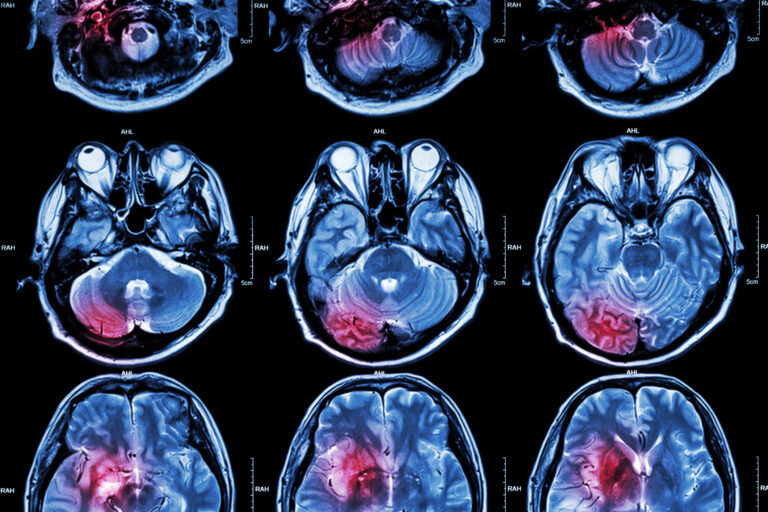
Magnetic resonance imaging (MRI) revolutionized medical diagnostics by evolving from nuclear magnetic resonance discoveries to life-saving technology.
Magnetic Resonance Spectroscopy (MRS), a variant of conventional Magnetic Resonance Imaging (MRI), is an advanced diagnostic technique that offers a unique glimpse into the molecular composition of tissues. Unlike standard MRI, which provides detailed images of anatomical structures, MRS focuses on the chemical contents of cells, making it a crucial tool in medical diagnostics and research.
This technique operates on the principle of nuclear magnetic resonance, detecting the presence and concentrations of various metabolites within the body. By analyzing these chemical signatures, MRS can identify subtle biochemical changes that often precede visible pathological alterations. This capability makes it particularly valuable for early diagnosis and for monitoring the progression of diseases, including cancer, neurological disorders, and metabolic dysfunctions.
One of the primary applications of spectroscopy MRI is in the field of neurology, where it is used to evaluate brain tumours, epilepsy, and degenerative diseases like Alzheimer’s and Parkinson’s. By measuring the levels of specific brain chemicals, such as N-acetylaspartate, choline, and creatine, clinicians can gain insights into cell density and brain metabolism, aiding in the assessment of disease severity and the response to therapy.
Furthermore, MRS is instrumental in oncology, where it helps differentiate between benign and malignant tumours based on their metabolic profiles. This differentiation is crucial for planning appropriate treatment strategies and predicting patient outcomes.
As research advances, the potential applications of spectroscopy MRI continue to expand, promising not only more precise diagnostic capabilities but also improved patient management and therapeutic outcomes. Through its non-invasive approach to revealing the biochemistry of diseases, spectroscopy MRI stands as a pivotal development in the fusion of imaging and molecular diagnostics.
home » Spectroscopy MRI
Magnetic resonance imaging (MRI) revolutionized medical diagnostics by evolving from nuclear magnetic resonance discoveries to life-saving technology.
