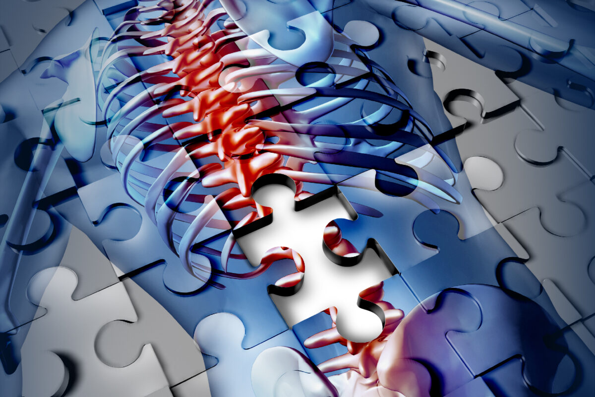
Revolution in the Spinal Cord: How New Imaging Tech is Transforming Diagnosis and Care
 By
By
Discover the latest developments in spinal cord imaging, including ultra-high-field MRI and artificial intelligence enhancements.
By
Discover the latest developments in spinal cord imaging, including ultra-high-field MRI and artificial intelligence enhancements.
 By
By
Uncover the impact of new MRI contrast agents on diagnostic imaging and their significance in modern medical technology.
 By
By
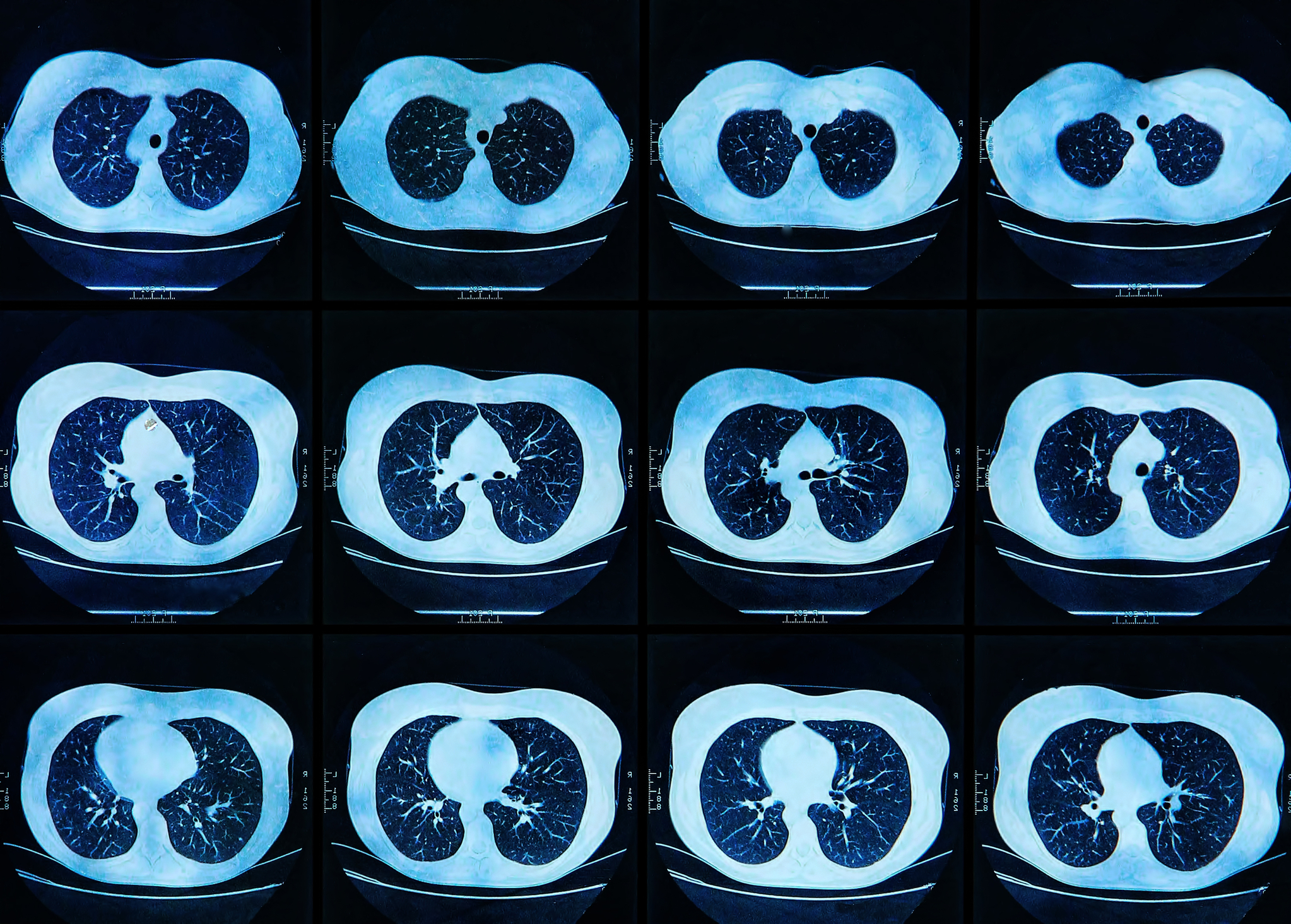
Discover how AI-powered low-field MRI is transforming lung imaging, enhancing accessibility and affordability for better healthcare.
 By
By
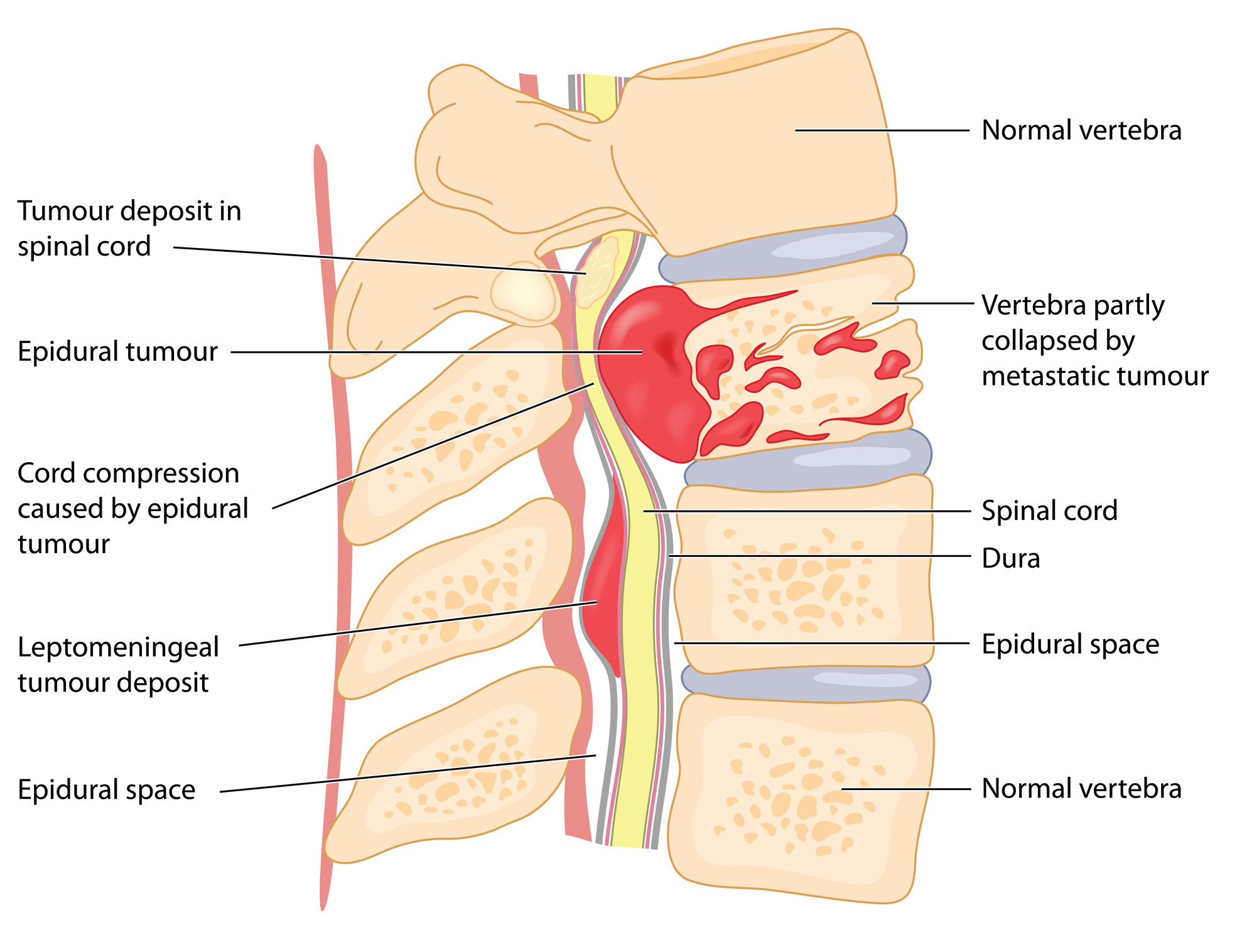
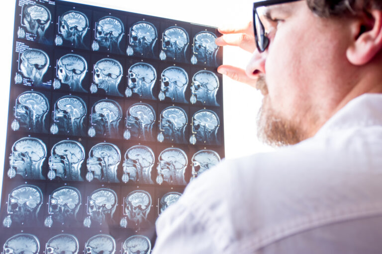
Discover the principles of MRI in spinal cord tumour diagnosis and how it helps identify various lesion types for improved care.
 By
By
Uncover the benefits of advanced MRI sequences in multiple sclerosis, offering deeper insights into disease mechanisms and treatments. Image for illustration only. Person depicted is a model.
 By
By
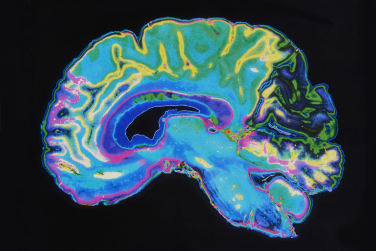
Discover the principles behind magnetic resonance theory and how it has advanced MRI as a vital tool in modern diagnostic imaging.
 By
By
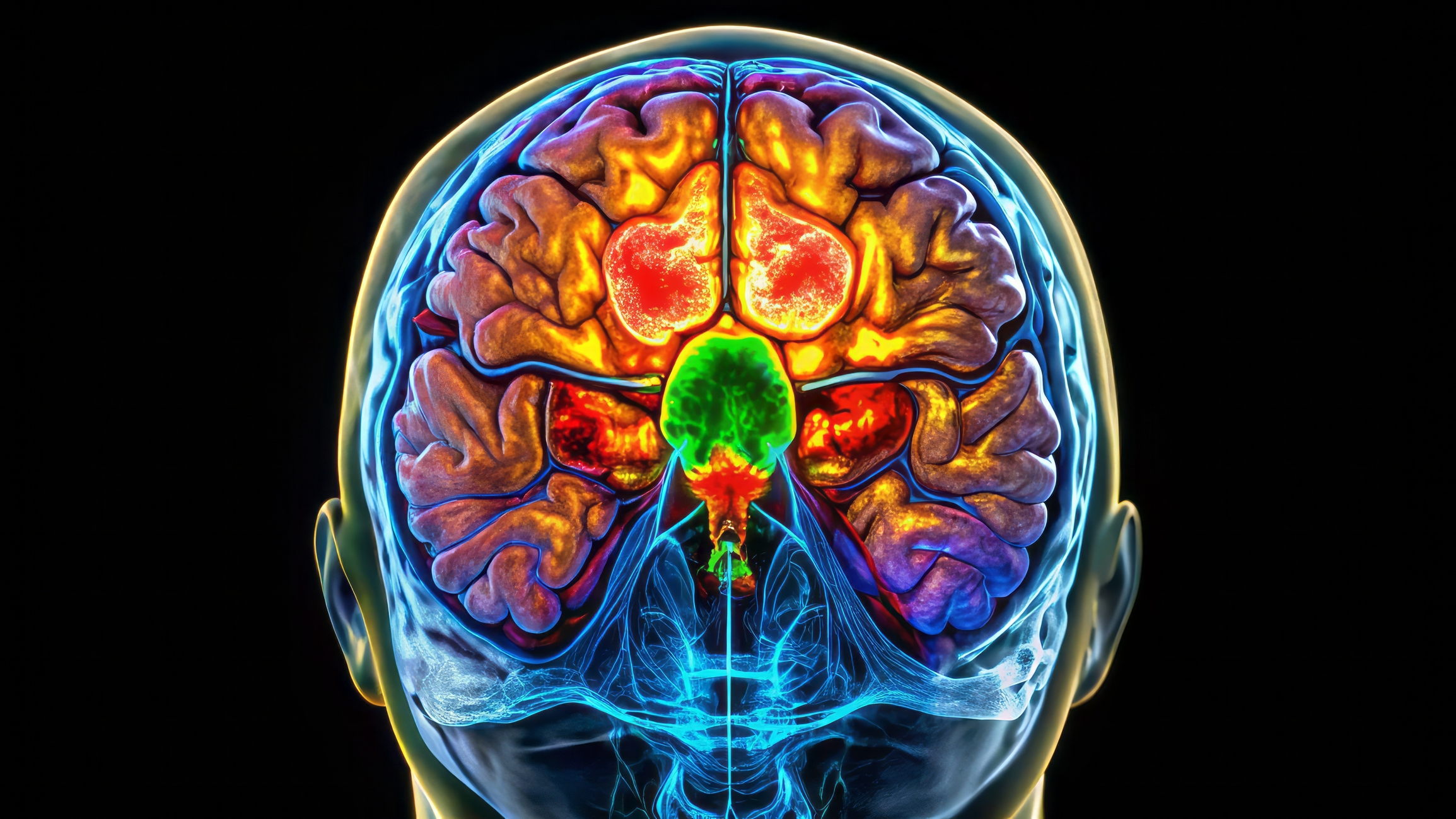
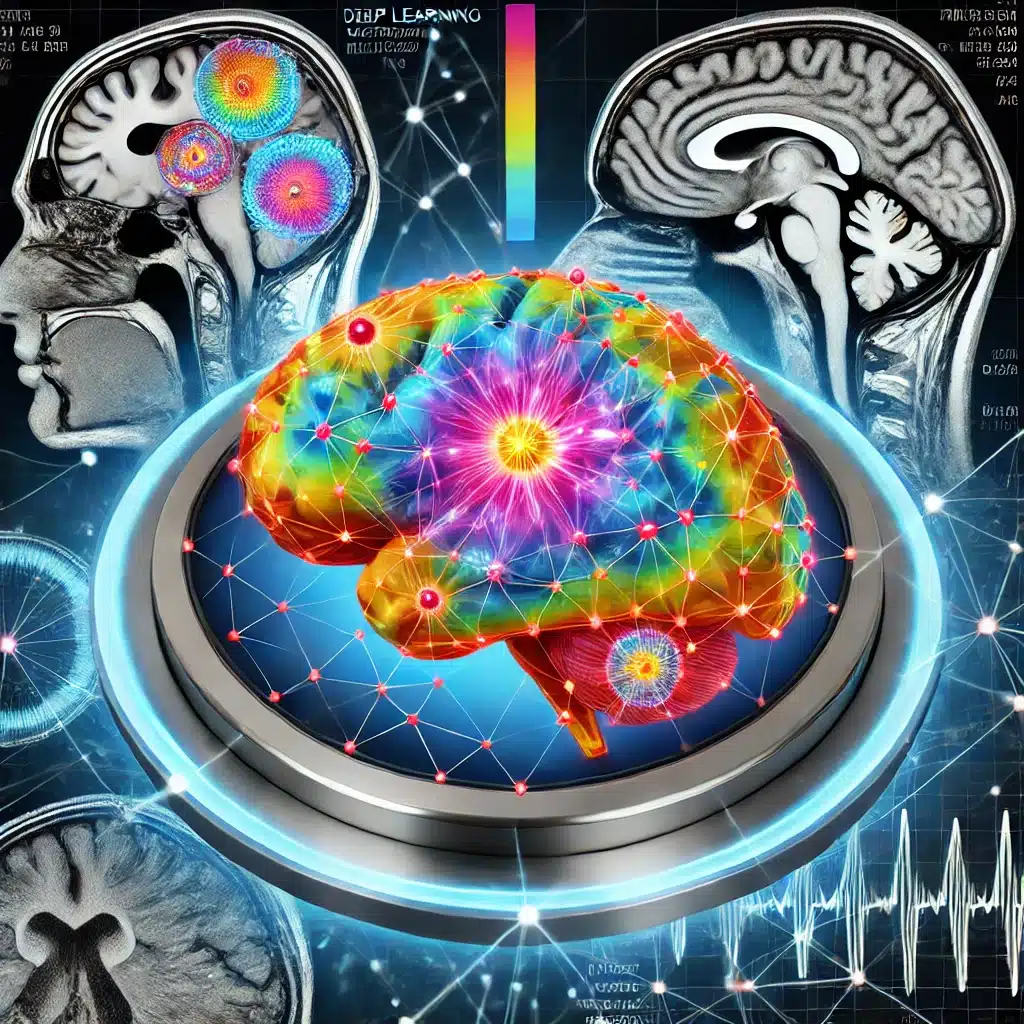
Stathopoulos et al. demonstrate that combining deep learning with MRI sequences significantly improves brain tumour detection, achieving up to 98.4% accuracy.
 By
By
Basics of MRI provide essential knowledge for understanding advanced imaging techniques, revolutionising medical diagnostics and patient care globally. Image for illustration only. Person depicted is a model.
 By
By
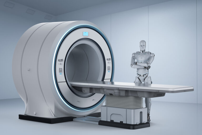
AI in MRI transforms medical imaging by increasing diagnostic accuracy, lowering costs, and broadening patient accessibility significantly.
 By
By
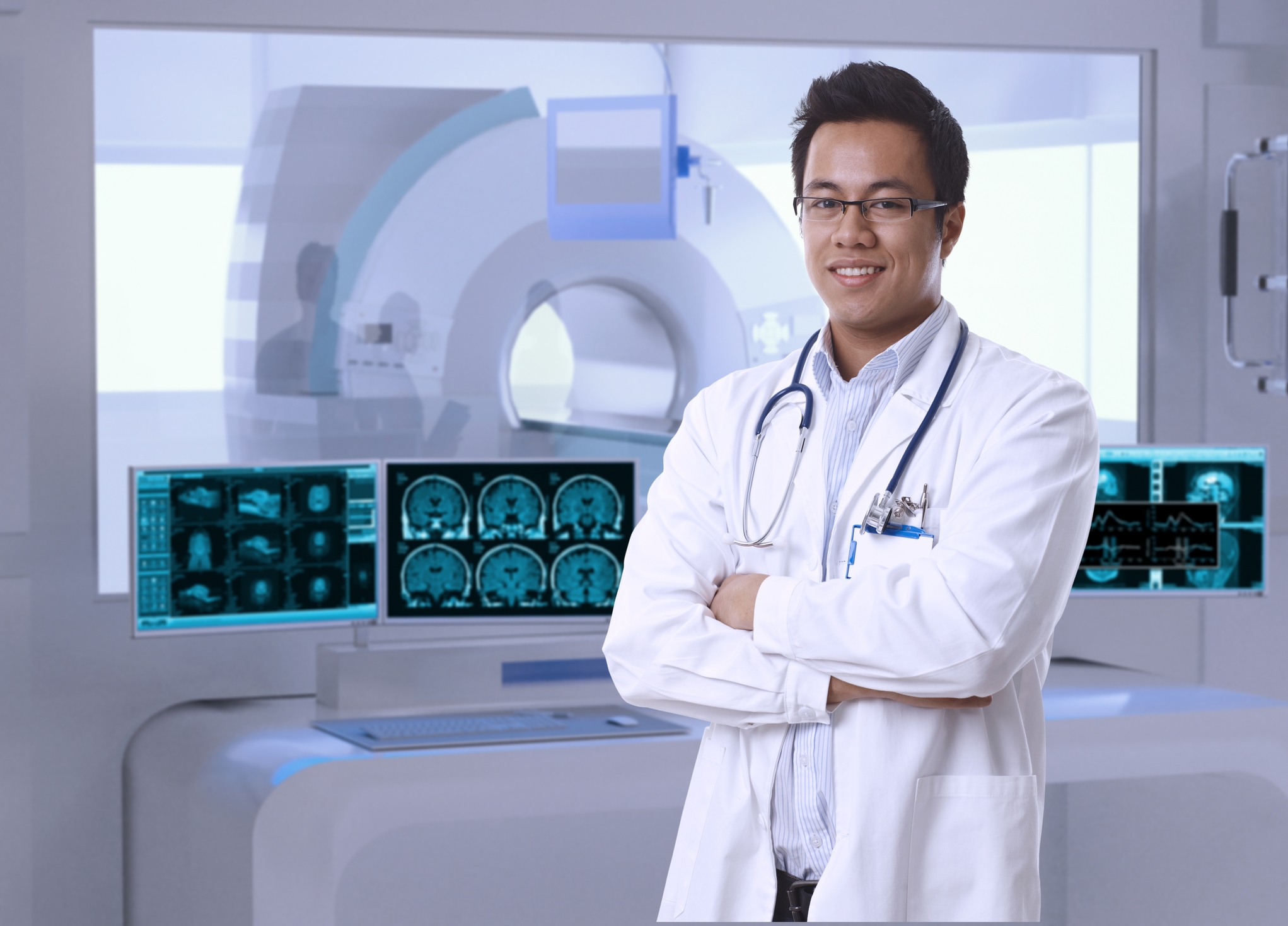
MRI scans provide critical insights into the body’s internal structures, aiding in the diagnosis and treatment of various conditions.
 By
By
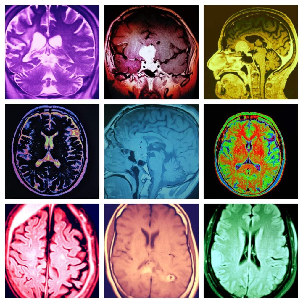
Understanding the different types of MRI involves exploring specialised techniques like fMRI, dMRI, MRS, and CMR, each offering unique diagnostic insights.