Ultrasound Elastography for Surgery
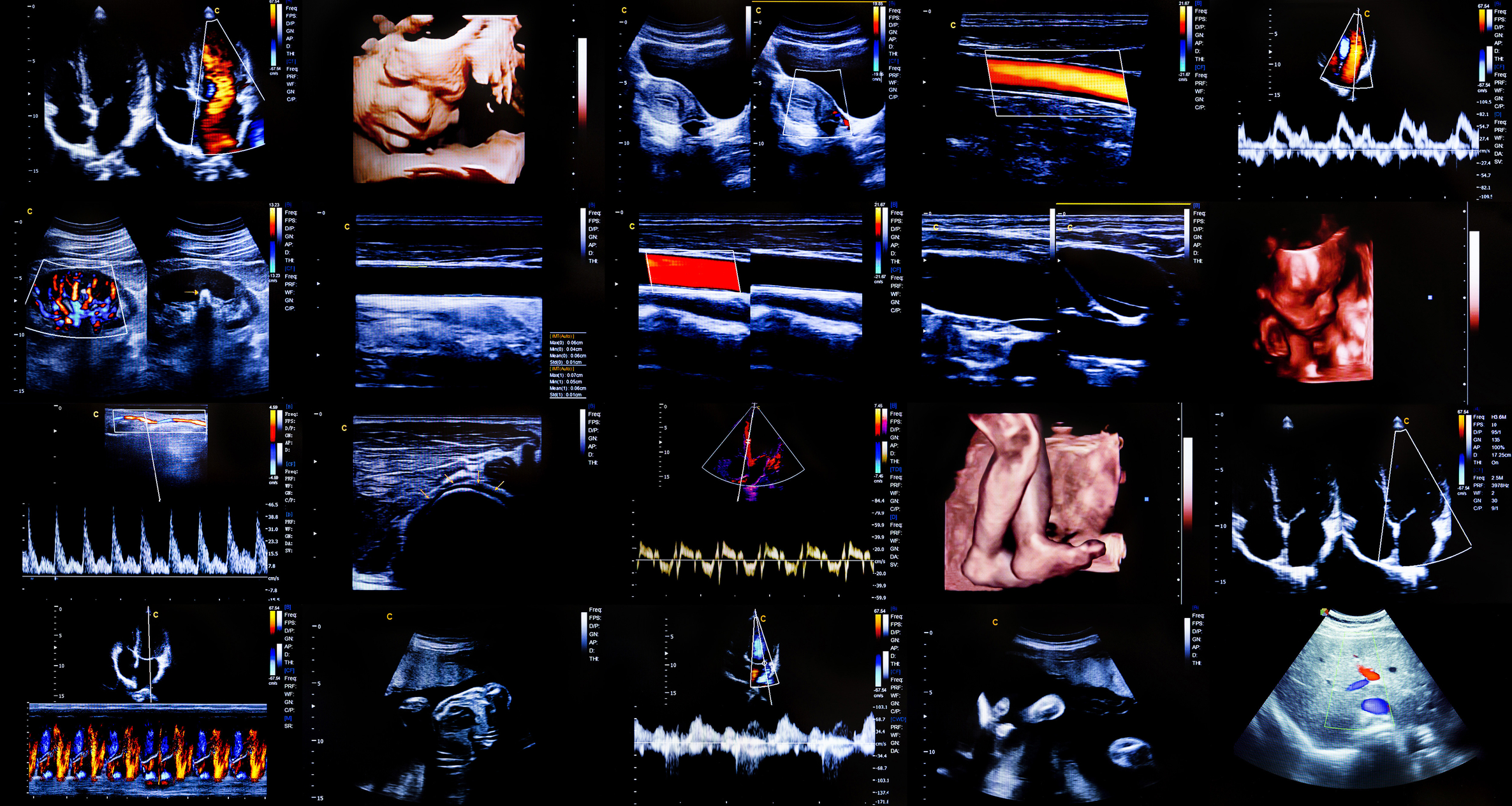
Ultrasound elastography has become an important tool in surgical planning and intraoperative decision-making. This imaging technique provides real-time information about tissue stiffness, aiding surgeons in distinguishing between normal and pathological tissues. Its non-invasive nature and ability to enhance surgical precision make it particularly valuable in procedures involving the liver, breast, thyroid, prostate, and musculoskeletal system.
Principles of Ultrasound Elastography for Surgery
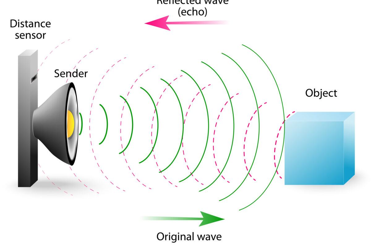
Elastography assesses tissue stiffness by measuring how tissues deform in response to mechanical stress. There are two main types: strain elastography and shear wave elastography (SWE).
- Strain Elastography involves applying external pressure (manual or physiological) to generate tissue displacement, which is then analysed to produce a stiffness map. This method is qualitative and highly dependent on operator skill.
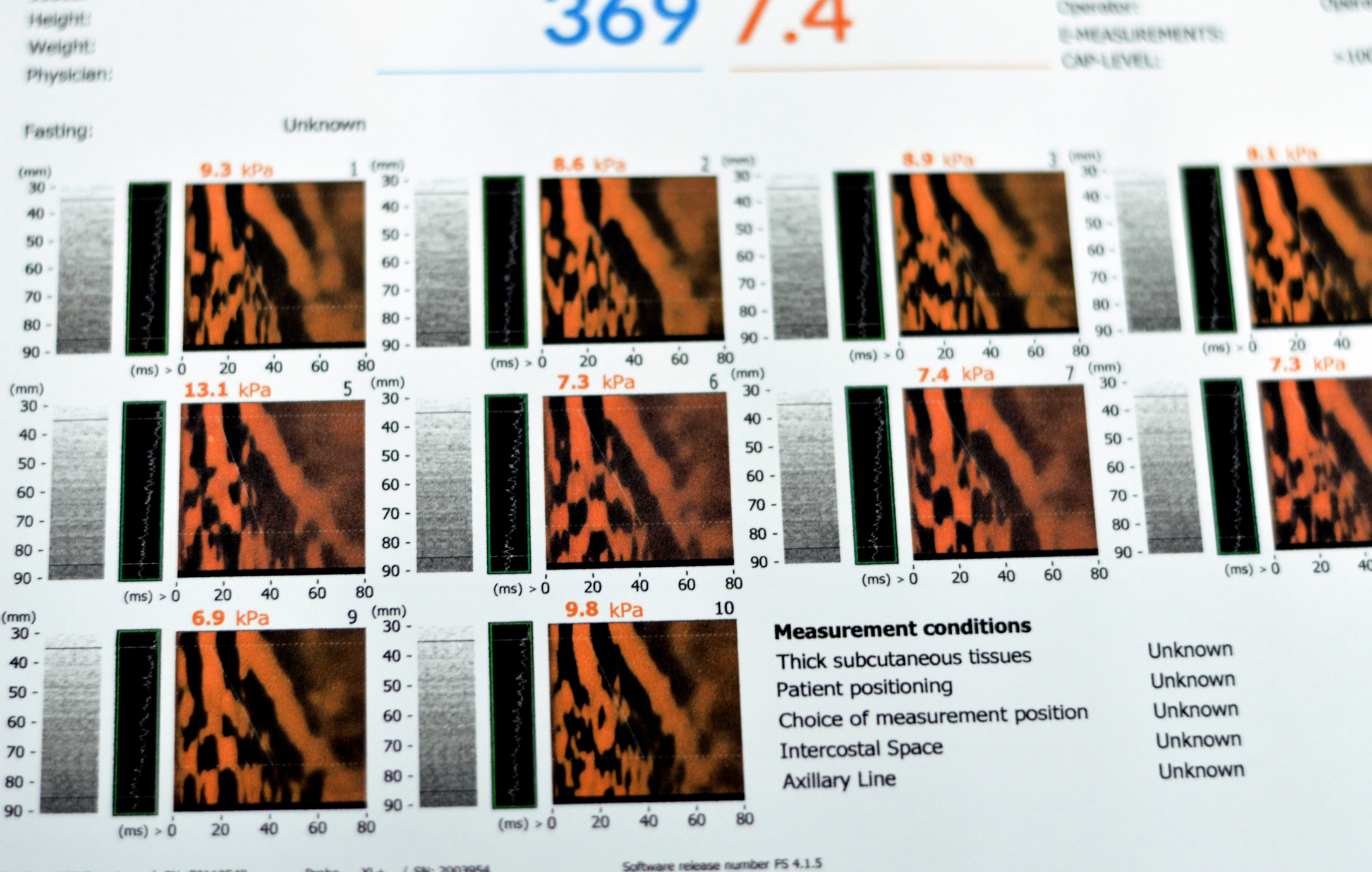
- Shear Wave Elastography uses acoustic radiation force to create shear waves, measuring their propagation speed to quantify tissue stiffness. This technique provides reproducible and objective data, making it particularly useful in clinical settings.
Applications in Surgery
- Oncological Surgery
In tumour resection, elastography helps differentiate between malignant and benign tissues. Many cancers exhibit increased stiffness due to fibrosis and desmoplastic reactions. Breast cancer, for instance, is often harder than surrounding tissue, making elastography useful for guiding biopsies and assessing tumour margins intraoperatively. Liver elastography assists in evaluating fibrosis severity, influencing decisions on surgical resectability and liver transplant eligibility.
- Thyroid and Parathyroid Surgery
Elastography improves preoperative planning by identifying malignant thyroid nodules, reducing unnecessary surgeries. Parathyroid adenomas, which are softer than surrounding tissues, can be distinguished more easily, aiding in minimally invasive parathyroidectomy.
- Prostate and Urological Surgery
In prostate cancer surgery, elastography assists in targeted biopsies, reducing false negatives. It also helps preserve neurovascular bundles by differentiating between tumour infiltration and normal tissue.
- Musculoskeletal Surgery
Elastography is useful in tendon and ligament surgery by assessing tissue integrity and guiding repair strategies. It helps evaluate muscle stiffness post-injury, influencing rehabilitation and surgical decisions.
Intraoperative Use
During surgery, elastography can be integrated with conventional ultrasound to provide real-time feedback on tissue characteristics. This is particularly beneficial in brain tumour surgery, where distinguishing between infiltrative tumour and normal brain tissue is crucial. Elastography’s ability to highlight differences in stiffness can improve resection completeness while preserving functional areas.
Limitations and Future Developments
While promising, elastography has limitations, including dependency on probe pressure in strain elastography and challenges in deep tissue imaging. Future advancements may focus on integrating artificial intelligence for automated analysis and improving real-time elastography-guided robotic surgery.
Ultrasound elastography continues to refine surgical precision, offering an additional layer of information that enhances both preoperative planning and intraoperative guidance. Its expanding role in various surgical fields highlights its growing importance in modern medical practice.
home » Ultrasound Elastography for Surgery
 By
By