Dental care has changed quietly but profoundly over the past few decades. The hand mirror and exploratory probe still exist, but they no longer carry the diagnostic burden alone.
Imaging now sits at the center of modern dental decision-making, shaping everything from routine checkups to complex surgical planning. It is not simply a supporting tool; in many cases, it is the starting point.
What makes dental imaging distinctive is its proximity to action. A scan is often followed by an immediate clinical decision: monitor, restore, extract, refer, or intervene surgically. Unlike some medical fields where imaging findings may sit in a chart for weeks, dental imaging is tightly woven into real-time care.
Seeing What the Eye Cannot
Here’s why imaging became indispensable in dentistry. Teeth and supporting structures hide their most important information beneath enamel, bone, and soft tissue. Pain, swelling, or discoloration may hint at a problem, but imaging reveals its true extent.
Modern dental imaging does not replace clinical judgment. It sharpens it.
Early Detection and Preventive Care
Radiographs allow dentists to identify issues long before symptoms appear. Interproximal caries, early bone loss, periapical pathology, and impacted teeth often develop silently. Imaging turns prevention into something tangible rather than theoretical.
A patient who feels “fine” may still have measurable changes visible on imaging. Catching these early alters treatment trajectories, cost, and long-term outcomes.
Establishing Baselines Over Time
Dental imaging also creates continuity. Comparing current images with prior studies reveals patterns of progression or stability. Bone levels, root integrity, and restoration margins are easier to assess when change is documented visually.
This longitudinal perspective is particularly valuable in periodontal care and orthodontics, where small shifts matter.
Imaging Modalities in Dental Practice
Dental imaging is not a single technique. Each modality answers different questions, and modern practice often combines several to form a complete picture.
Intraoral Radiographs and Their Precision
Intraoral radiographs remain foundational. Bitewings, periapical images, and occlusal views provide high-resolution detail of individual teeth and surrounding bone. They are quick, targeted, and effective for diagnosing decay, abscesses, and root morphology.
Despite advances in three-dimensional imaging, these two-dimensional views remain irreplaceable for routine diagnostics.
Panoramic Imaging and the Broader View
Panoramic imaging offers context. It captures the jaws, teeth, temporomandibular joints, and surrounding structures in a single image. This broader perspective is essential for evaluating tooth eruption patterns, jaw asymmetry, cysts, and developmental anomalies.
Panoramic scans often serve as the first step in assessing complex cases, especially when surgical planning is anticipated.
Cone Beam CT and Three-Dimensional Insight
Cone beam computed tomography (CBCT) has reshaped dental imaging. It provides three-dimensional views of teeth, bone, nerves, and sinus anatomy with a level of detail previously unavailable in dental settings.
CBCT is particularly valuable for implant planning, endodontic assessment, and oral surgery. It reduces uncertainty by allowing clinicians to measure distances, assess bone quality, and identify anatomical variations before intervention.
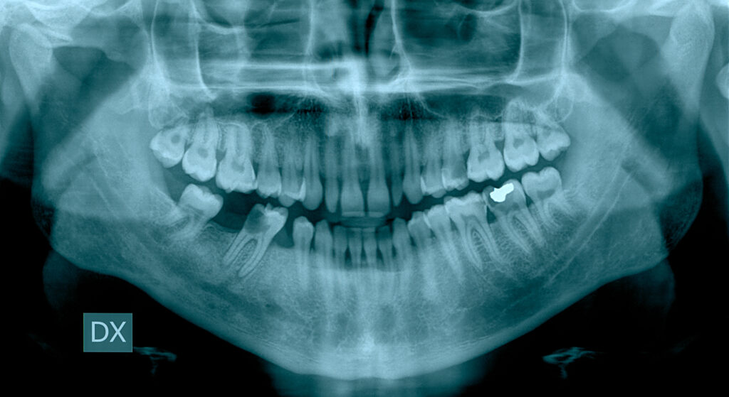
Imaging and Wisdom Teeth: A Practical Intersection
Few areas of dentistry illustrate the value of imaging as clearly as wisdom teeth management. Third molars vary widely in position, eruption pattern, and proximity to critical structures. Visual inspection alone is rarely sufficient.
Understanding Position, Angulation, and Risk
Imaging reveals whether wisdom teeth are fully erupted, partially impacted, or completely embedded in bone. Angulation—horizontal, mesioangular, vertical, or distoangular—directly influences the risk of future complications and the complexity of removal.
Proximity to the inferior alveolar nerve or maxillary sinus is another critical consideration. These relationships are often invisible without imaging, yet they significantly affect surgical planning and patient counseling.
When Monitoring Is Better Than Removal
Not all wisdom teeth require extraction. Imaging helps differentiate between teeth that pose an active risk and those that can be safely monitored. Bone coverage, follicular space size, and adjacent tooth health guide this decision.
For a deeper guidance on this topic, See NSOMS for more info about wisdom teeth and removal.
Imaging as a Surgical Safety Tool
When removal is indicated, imaging reduces uncertainty. Knowing nerve position, root shape, and bone density allows for more precise, conservative surgery. This translates into fewer complications, shorter recovery, and clearer informed consent.
In wisdom tooth care, imaging is not optional—it is protective.
Imaging in Restorative and Prosthetic Dentistry
Beyond diagnosis and surgery, imaging supports restorative accuracy. Crowns, bridges, and implants depend on precise measurements and anatomical awareness.
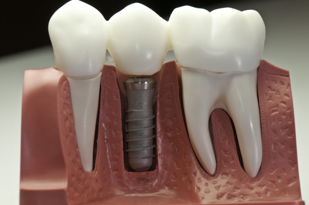
Implant Planning and Bone Assessment
Dental implants require adequate bone volume and favorable anatomy. CBCT imaging allows clinicians to evaluate bone height, width, and density before placement. It also identifies sinus boundaries and nerve canals that must be respected.
Imaging-guided planning improves implant longevity and reduces the risk of failure.
Evaluating Existing Restorations
Imaging also helps assess the integrity of existing work. Recurrent decay under restorations, marginal gaps, and root fractures may not be visible clinically. Radiographs provide clarity when symptoms and surface findings do not align.
Periodontal and Endodontic Applications
Imaging plays a central role in both periodontal and endodontic care, where disease often progresses below the gum line.
Bone Loss and Periodontal Mapping
Radiographs document bone levels around teeth, revealing patterns of periodontal disease. Vertical and horizontal bone loss, furcation involvement, and progression over time become visible through imaging.
This information guides treatment planning and helps patients understand disease severity.
Root Canal Anatomy and Complexity
Endodontic treatment relies heavily on imaging. Root curvature, canal number, and anatomical variations determine treatment difficulty. CBCT imaging can reveal additional canals or fractures that standard radiographs may miss.
Accurate imaging reduces retreatment rates and procedural complications.
Patient Communication and Trust
One often overlooked role of imaging in dental practice is communication. Images make abstract explanations concrete.
Visualizing the Problem
Patients may struggle to understand why a procedure is recommended when pain is minimal or absent. Showing an image of decay approaching the pulp or bone loss around a tooth bridges that gap.
Seeing creates understanding, and understanding improves compliance.
Supporting Informed Consent
Imaging also supports ethical care. When patients can see the structures involved and the risks present, consent becomes meaningful rather than procedural. This is particularly important in surgical decisions, including wisdom tooth removal and implant placement.
Balancing Technology With Judgment
While imaging is powerful, it is not infallible. Artifacts, positioning errors, and incidental findings can mislead if interpreted without context. Modern dental practice requires balance.
Imaging should inform decisions, not dictate them in isolation. Clinical examination, patient history, and symptoms remain essential.
The Future of Dental Imaging
As technology advances, imaging will continue to integrate more deeply into dental workflows. Artificial intelligence, enhanced resolution, and lower radiation doses are already shaping the next phase.
What will not change is the role imaging plays as a bridge between observation and action. It allows dentists to move from guesswork to precision, from reactive care to proactive planning.
In modern dental practice, imaging is not just about seeing teeth. It is about understanding the structures, risks, and possibilities that lie beneath the surface—and using that understanding to deliver care that is safer, clearer, and more effective for every patient.
Disclaimer
This article is published for general information and educational purposes only. It does not constitute dental, medical, surgical, or professional advice, nor is it intended to replace consultation with a qualified dental or healthcare professional. The content reflects general principles and practices within modern dentistry at the time of publication and may not apply to every individual case or clinical situation.
Diagnostic imaging techniques, treatment decisions, and clinical outcomes vary depending on patient-specific factors, practitioner judgement, available technology, and local regulatory guidance. Readers should not act upon any information contained in this article without seeking appropriate professional advice tailored to their individual circumstances.
Open MedScience does not accept responsibility for any loss, harm, or adverse outcomes arising from the use or interpretation of the information presented. References to external organisations or resources are provided for informational purposes only and do not imply endorsement.
You are here: home » diagnostic medical imaging blog »