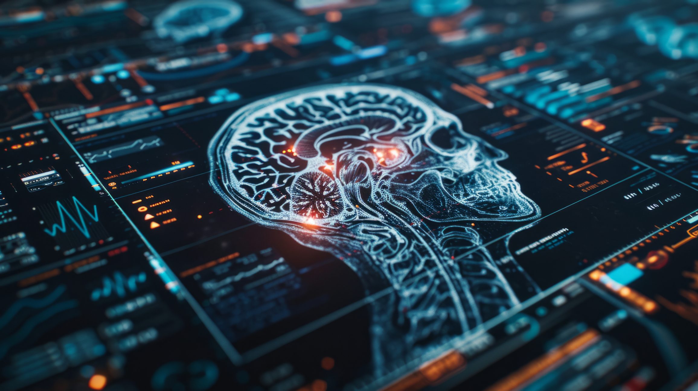
Neuroimaging is entering an era of accelerated progress, driven by innovations in magnetic resonance technology, advances in computational science, and a growing understanding of neurological and psychiatric disorders. These developments are redefining how researchers study the brain, how clinicians diagnose disease and how therapies are personalised. The current wave of progress spans structural imaging, functional mapping, machine learning, vascular imaging, and early-stage disease detection. This article explores some of the most influential trends shaping the field today and how they may transform neuroscience and clinical practice.
Personalised neuromodulation guided by advanced imaging
One of the most striking shifts in neuroimaging is the rapid integration of imaging data with neuromodulation techniques. Transcranial Magnetic Stimulation (TMS) has been used for years to treat conditions such as depression and anxiety, yet its effectiveness has varied from person to person. A significant factor in this variation is the diversity of individual brain structures and networks. New imaging-led approaches are beginning to address this limitation.
Modern functional MRI, diffusion imaging and high-resolution structural scans now enable researchers to create detailed maps of each person’s brain. These maps reveal connectivity patterns, cortical thickness variations and individualised network signatures. When combined with machine learning methods, they enable clinicians to identify more precise TMS stimulation targets. This approach shifts treatment planning from generic guidance based on standard anatomical landmarks to truly personalised targeting.
Such precision mapping offers several benefits. Treatment may become more consistent and predictable simply because the stimulation is delivered to regions verified as relevant to the person’s specific symptoms. Interventions can also be adapted over time if new imaging data show changes in network connectivity. Challenges remain, including the time and expense required for high-quality imaging and the need for robust clinical validation. Even so, this integration of imaging and neuromodulation is one of the most evident signs of where therapeutics are heading.
Machine learning and predictive models in vascular and neurodegenerative imaging
Across the broader field of medical imaging, machine learning is increasingly influencing how diseases are identified and monitored, particularly in neuroimaging. One area receiving significant attention involves cerebral small vessel disease (CSVD), which contributes to vascular dementia and cognitive decline. Traditionally, clinicians have relied on visual assessment of MRI markers such as white-matter hyperintensities, microbleeds and lacunes. These assessments can be subjective and vary across observers.
Machine learning models offer a different approach. They automatically detect, measure, and classify lesions and can integrate multiple imaging markers to produce predictions about cognitive trajectory, disease severity, or the risk of conversion to dementia. Such models can handle subtle patterns that may be difficult for humans to perceive and produce combined metrics that capture risk in new ways.
Another rapidly growing application is “brain age” estimation. By training algorithms on large datasets of MRI scans, researchers can predict a brain’s biological age. When this biological age differs significantly from a person’s chronological age, the gap may signal early neurodegenerative change. Brain-age models have been investigated for Alzheimer’s disease, frontotemporal dementia and other conditions where early detection is essential.
These systems are still in development and must be validated across different scanners, populations and clinical environments. Still, the direction of travel is clear: neuroimaging is becoming more predictive rather than simply descriptive, with algorithms offering early warning signs long before symptoms appear.
Imaging the prodromal phase of Parkinson’s disease
Parkinson’s disease has traditionally been diagnosed based on motor symptoms such as tremor, rigidity and bradykinesia. However, growing evidence suggests that the disease starts many years before these symptoms emerge. Non-motor signs such as REM sleep behaviour disorder, reduced sense of smell, and subtle cognitive changes may appear in the prodromal phase, long before clinical diagnosis.
Neuroimaging has started to play a central role in understanding this early phase. Researchers are exploring structural MRI, functional MRI, diffusion imaging and molecular imaging to reveal changes in brain regions involved in olfaction, sleep regulation and dopamine pathways. High-resolution imaging of the substantia nigra, in particular, has shown promise for detecting microstructural alterations that precede the onset of motor symptoms.
The possibility of imaging-based early detection raises essential questions. If clinicians can identify people at higher risk long before symptoms develop, therapeutic interventions could potentially start earlier. Drug studies could also focus on disease-modifying treatments at a point where neuronal loss is not yet severe. Nonetheless, widespread screening is not yet practical, and markers must be accurate enough to avoid misdiagnosis. Even so, the growing emphasis on the prodromal period indicates a shift towards prevention rather than reaction.
The expanding role of neuropsychiatric and multisensory imaging
Neuropsychiatric symptoms in conditions such as dementia have long been challenging to characterise. Behavioural changes, agitation, apathy and affective disturbances often emerge early in the disease process, yet their neural underpinnings are not always clear. New imaging work is beginning to illuminate these relationships.
Multimodal imaging studies are mapping the networks associated with neuropsychiatric symptoms, linking them to patterns of structural atrophy, connectivity disruption or altered functional activation. Instead of treating behavioural symptoms as secondary complications, researchers now view them as integral parts of the disease process. This shift could lead to more targeted therapeutic approaches that focus on affected networks rather than general symptom management.
A further interesting development concerns multisensory processing. Imaging investigations of taste, chewing, swallowing and associated sensory integration have expanded significantly. These studies help explain how oral functions are coordinated and how neurological conditions disrupt them. The work bridges neuroscience, nutrition, dental health and speech therapy, creating opportunities for cross-disciplinary collaborations.
There is also continued interest in how large-scale stress events influence the brain. Studies examining the long-term effects of the COVID-19 pandemic on neural function have revealed structural and functional changes in regions associated with anxiety, stress regulation and memory. These findings suggest that neuroimaging can contribute to public health strategies, not only by assessing disease but also by informing mental health interventions.
Vascular imaging and advances in stroke and aneurysm assessment
Acute stroke care has always relied heavily on imaging, but recent innovations have refined how clinicians assess which tissues are salvageable and how quickly interventions must be delivered. Improvements in collateral-flow imaging allow doctors to evaluate whether blood flow around a blocked vessel is sufficient to sustain brain tissue until reperfusion can be attempted. These insights help determine who may benefit from thrombectomy or thrombolysis, even several hours after symptom onset.
In parallel, imaging of intracranial aneurysms continues to improve. Enhanced CTA and MRA techniques provide more precise visualisation of aneurysm structure, wall characteristics and rupture risk. Digital subtraction angiography remains the gold standard for high-detail imaging, but new processing techniques and computational modelling are making it easier to integrate imaging into decision-making. For interventional radiologists and neurosurgeons, these developments support more accurate assessment, improved procedural planning and safer treatment.
Such advances benefit not only specialist centres but also broader emergency-care networks. Faster imaging, automated triage tools and standardised protocols help ensure that patients receive the correct treatment pathway from the moment they enter the system.
The rise of computational and multimodal integration
Perhaps the most transformative movement across neuroimaging is the shift towards computational integration. Instead of evaluating individual scans in isolation, researchers are combining structural MRI, functional MRI, diffusion data, molecular imaging, electrophysiology, genetics and clinical scores into unified analytical frameworks. The aim is to understand the brain as a connected system and to identify patterns that emerge only when multiple data types are analysed together.
This shift is supported by growing access to large datasets, improved computational power and expanding open-science initiatives. Multimodal data integration helps uncover subtle network changes involved in psychiatric conditions, neurodegenerative disease and developmental disorders. It also supports efforts to personalise treatment by identifying markers specific to individuals rather than groups.
With this growth comes challenges. Different imaging centres use different scanners, protocols and processing tools, making standardisation difficult. Machine learning methods can be hard to interpret, leaving clinicians unsure how best to apply algorithmic findings. The sheer volume of data requires new storage and processing strategies. Nevertheless, computational neuroimaging is now central to the field’s evolution.
Looking ahead
The recent developments across neuroimaging point to a future where diagnosis is earlier, treatment is tailored, and research is more integrated than ever before. Personalised neuromodulation may become routine rather than exceptional. Machine learning tools could help clinicians predict disease years before symptoms appear. Multimodal datasets may reveal the pathways that underlie complex neuropsychiatric symptoms. Advances in vascular imaging will continue to improve outcomes in emergency settings, and high-resolution techniques may identify disease markers that were previously invisible.
However, successful translation of these developments into clinical practice requires careful validation, standardisation across centres and strong collaboration between scientists, clinicians and computational experts. Ethical considerations around data use, algorithmic fairness and access to advanced technologies must also be addressed.
Neuroimaging is entering a new era, driven by technology, computation and cross-disciplinary insight. As these innovations mature, they hold the promise of reshaping our understanding of the brain and improving the lives of patients living with neurological and psychiatric conditions.
Disclaimer
This article is provided for informational and educational purposes only. It summarises current research directions and technological developments in neuroimaging, but it is not intended to offer medical advice, clinical guidance or recommendations for patient care. The technologies, methods and applications discussed may still be under investigation, subject to regulatory oversight, or limited to research environments. Clinical decisions should always be made by qualified healthcare professionals based on individual circumstances, established guidelines and validated evidence.
Open MedScience does not endorse any specific technique, diagnostic tool, treatment or research approach mentioned in this article. While every effort has been made to ensure accuracy at the time of publication, scientific understanding evolves quickly, and interpretations may change as new findings emerge. Open MedScience accepts no liability for any actions taken based on the information presented.
home » blog » medical technologies »


