Automated cell counters are transforming medical imaging by enhancing accuracy and efficiency. These technologies are vital in delivering precise diagnostic outcomes. This article examines their role and impact in modern radiology.
Precision in medical imaging is crucial for accurate diagnostics, where even small errors can have significant consequences. Advanced cell analysis technologies have become important tools, ensuring imaging processes are both accurate and efficient. An accurate cell counter provides precise measurements essential for effective diagnostics. As these technologies evolve, their integration into medical imaging promises to redefine diagnostic accuracy standards.
Integration of cell analysis in imaging processes
Cell analysis technologies naturally complement medical imaging processes, offering unmatched precision. These systems integrate into existing workflows, allowing radiologists to gain deeper insights without disrupting established practices. The use of advanced cell analysis enhances diagnostic accuracy by providing detailed cellular-level information that traditional methods might miss. This integration ensures radiology labs consistently deliver precise diagnostic outcomes.
Moreover, these technologies improve diagnostic precision by minimising human error. Automated systems ensure consistent results, which is crucial for maintaining high standards of patient care. The ability to analyse cellular data quickly and accurately empowers healthcare providers to make informed decisions promptly. Such advancements are invaluable in fast-paced medical environments where timely diagnosis can significantly impact patient outcomes.
Advancements in technology enhance imaging accuracy
Cell analysis has seen remarkable technological advancements that significantly contribute to the accuracy of medical imaging. Innovations such as enhanced sensor technology and sophisticated algorithms enable more detailed and reliable cellular data analysis. These developments improve the efficiency of imaging processes and elevate the quality of diagnostic information obtained.
For instance, automated cell counters now employ advanced optical or impedance-based methods to enumerate and characterise cells with high precision, as highlighted in this study on automated cell counting. Concurrently, deep-learning and machine-learning models are increasingly applied to cell image classification (e.g., blood-cell subtypes, viability assessments), reducing subjective interpretation and increasing reproducibility, as described in research on AI in medical imaging. These technological strides have enabled radiology labs to achieve higher precision levels in their diagnostic capabilities, allowing for earlier detection and better disease management.
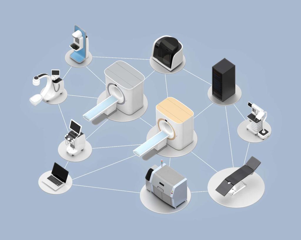
Integration of cell-analysis outputs with radiology information systems and AI-supported imaging workflows means the data can be used in real time to flag anomalies or suggest further investigation. For example, an automated cell counter detecting an unexpectedly high count of a particular cell type could signal to the radiologist to focus on a specific region or parameter. This synergy fosters a smarter, more responsive diagnostic environment.
Impact on radiology labs and diagnostic improvements
Incorporating advanced cell analysis into radiology labs has markedly improved diagnostic outcomes, setting new benchmarks for precision in medical imaging. Radiologists now have access to tools that offer enhanced detail and clarity, contributing to more accurate interpretations and diagnoses. The integration of these technologies has streamlined processes and significantly reduced the margin for error.
With these enhancements, labs report improved efficiency and effectiveness in diagnosing complex conditions. The ability to rely on robust data analysis ensures healthcare providers consistently deliver high‑quality patient care. As a result, patients benefit from quicker diagnosis and treatment plans tailored to their specific needs.
Importantly, the combined workflow of imaging and cellular analysis reduces the need for multiple repeat scans or biopsies, thereby lowering patient risk, reducing costs, and speeding up decision-making. By giving clinicians a more complete picture (from cellular detail to whole‑body imaging), the treatment pathway becomes smoother and more personalised.
Future trends in cell analysis technology
The future of cell analysis technology holds great promise for further transforming medical imaging. Emerging trends suggest a continued focus on automation and artificial intelligence integration, enhancing both speed and accuracy in diagnostics. These innovations are expected to provide more personalised insights into patient conditions, leading to tailored treatment approaches.
As technology advances, the potential for groundbreaking improvements in diagnostic processes grows exponentially. The ongoing evolution of cell analysis technology will likely lead to more refined imaging techniques, offering unparalleled detail and precision. This continuous innovation ensures that medical professionals remain equipped with cutting-edge tools necessary for delivering optimal patient care.
Looking ahead, we can expect more seamless integration of cell analysis with multi-modality imaging, real-time monitoring in critical care settings, and point-of-care devices capable of rapid cellular and imaging assessment at the bedside. In turn, this could revolutionise how radiologists and pathologists collaborate, bridging cellular pathology with in vivo imaging. Scientific literature supports these advancements, highlighting the integration of cell analysis in medical imaging as a pivotal development.
Studies have demonstrated that automated cell counters significantly enhance diagnostic accuracy, reducing the likelihood of human error, as shown in this PMC article. Moreover, the application of artificial intelligence in cell analysis has been shown to improve the speed and reliability of diagnostic processes, as discussed in this BMC Medical Imaging study. These references underscore the transformative impact of advanced cell analysis technologies in modern radiology.
Disclaimer
The information presented in “Improving Medical Imaging with Advanced Cell Analysis Technology” by Open MedScience is intended for general educational and informational purposes only. It does not constitute professional medical advice, diagnosis, or treatment. Readers should not rely on the material as a substitute for consultation with qualified healthcare or scientific professionals.
While efforts have been made to ensure the accuracy and reliability of the content at the time of publication, Open MedScience makes no representations or warranties regarding the completeness, accuracy, or applicability of any information contained herein. The technologies and methods discussed may not be suitable for all clinical or research settings, and outcomes may vary depending on implementation and context.
Open MedScience does not endorse any specific products, manufacturers, or technologies mentioned in the article. References to studies or external sources are provided for informational purposes and do not imply endorsement or verification by Open MedScience. Readers are encouraged to consult peer-reviewed literature and relevant professionals before making decisions based on the information provided.
Open MedScience and its contributors disclaim any liability for any direct or indirect loss, injury, or damage arising from the use or reliance on the information contained in this publication.
home » blog » medical technologies »


