Medical Imaging Breakthroughs Driving the Future of Healthcare
 By
By
Stay updated on medical imaging breakthroughs that are transforming healthcare and improving patient outcomes in innovative ways.
Artificial Intelligence-based radiomics represents a transformative approach to medical imaging, combining radiological data with advanced computational techniques to enhance diagnosis, prognosis, and treatment monitoring. By extracting quantitative features from medical images, radiomics aims to uncover patterns and correlations that are not discernible through traditional visual analysis. When integrated with AI, these methods can process vast datasets and deliver insights with unprecedented accuracy and speed.
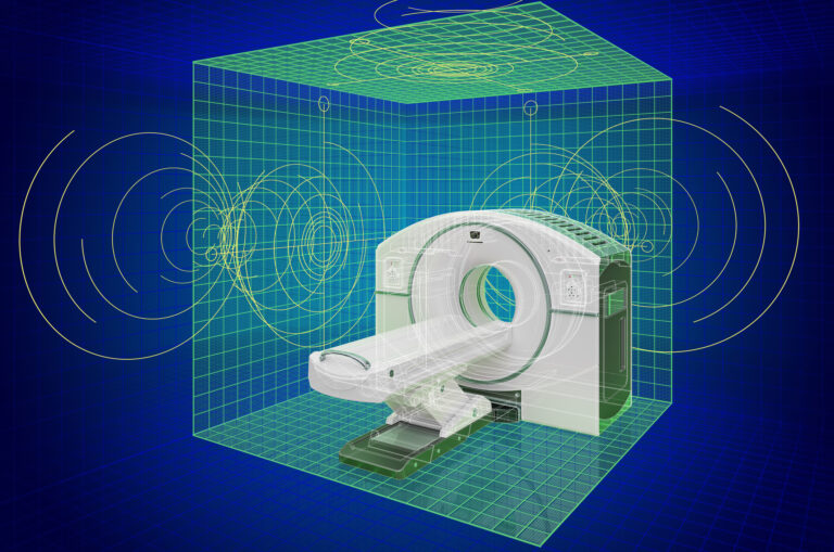
Radiomics begins with the acquisition of high-quality medical images, typically from modalities such as computed tomography (CT), magnetic resonance imaging (MRI), or positron emission tomography (PET). These images undergo pre-processing steps to standardise quality and resolution, ensuring consistency across data inputs. The next phase involves feature extraction, where mathematical algorithms quantify attributes like shape, texture, intensity, and wavelet transformations. These features capture the heterogeneity within a tumour or lesion, providing a rich dataset that reflects the underlying biology.
AI, particularly machine learning (ML) and deep learning (DL), plays a pivotal role in analysing the extracted features. ML algorithms, such as random forests or support vector machines, can identify complex relationships between radiomic features and clinical outcomes. Deep learning, on the other hand, employs neural networks to automatically learn feature representations from raw imaging data. These AI models are trained using annotated datasets, where outcomes such as disease progression or treatment response are known, enabling the system to make predictions on new, unseen cases.
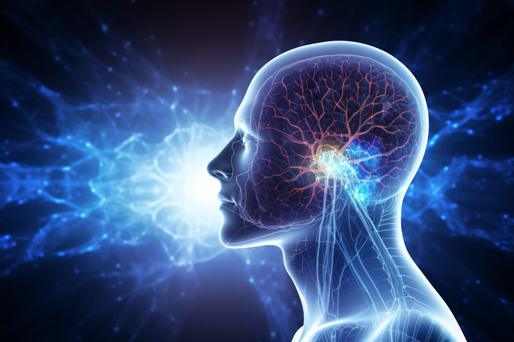
One of the most significant applications of AI-based radiomics is in oncology. It is increasingly used to characterise tumours, predict response to therapy, and assess treatment outcomes. For instance, AI models can differentiate between benign and malignant lesions with high sensitivity and specificity. They can also predict which patients are more likely to respond to specific treatments, allowing for personalised therapeutic strategies. Beyond oncology, AI-based radiomics has applications in neurology, cardiology, and respiratory medicine, offering similar advantages in terms of precision and predictive power.
Despite its potential, several challenges remain in implementing AI-based radiomics in clinical practice. Data standardisation is critical, as variations in imaging protocols can affect feature reproducibility. Additionally, the need for large, diverse datasets to train robust AI models poses logistical and ethical concerns, particularly regarding data privacy and sharing. Regulatory approval and clinician acceptance further influence the integration of these technologies into routine workflows.
In conclusion, Artificial Intelligence-based radiomics represents a promising frontier in medical imaging, capable of uncovering nuanced patterns that improve patient outcomes. While challenges exist, ongoing advancements in AI algorithms, imaging technologies, and data infrastructure are likely to overcome these barriers, paving the way for widespread adoption in precision medicine.
home »
By
Stay updated on medical imaging breakthroughs that are transforming healthcare and improving patient outcomes in innovative ways.
 By
By
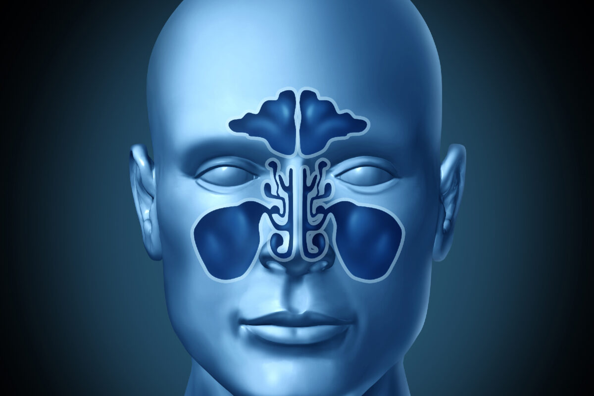
Discover how nasal and sinus imaging has evolved and its crucial role in diagnosing conditions like rhinosinusitis and tumours.
 By
By
Discover how medical imaging in 2025 has evolved with AI integration and population datasets shaping patient care.
 By
By
Explore total-body PET imaging and its advancements in sensitivity and clinical applications for better molecular insights.
 By
By
Learn about the advances in medical imaging that enhance diagnostics and patient experiences through AI and hybrid systems.
 By
By
Discover the impact of generative AI in diagnostics, enabling automated reporting and improving the efficiency of radiologists.
 By
By
Discover the role of AI in radiology automation, from scan triage to quality checks, transforming healthcare delivery today.
 By
By
Medical Science Trends in 2025 focus on AI, personalised treatments, and advancing roles for healthcare professionals.
 By
By
Digital Twin of the Heart creates a precise virtual model to predict, monitor, and improve cardiovascular health outcomes. Image for illustration only. Person depicted is a model.
 By
By
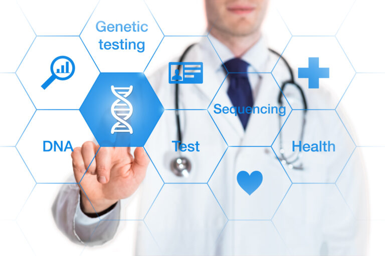
Ancestry DNA data helps uncover family roots, ethnic origins, health traits, and potential genetic connections globally. Image for illustration only. Person depicted is a model.