Cone Beam CT
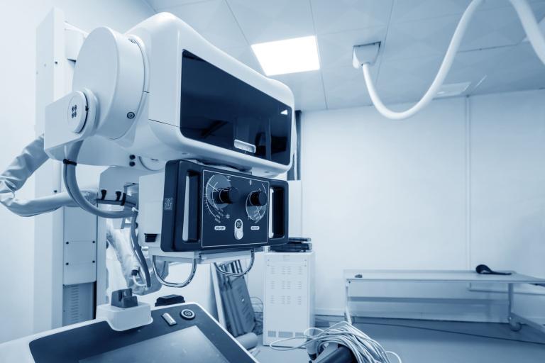
Cone Beam CT (CBCT) stands out in the field of medical imaging due to its unique ability to provide 3D images of the body, which is particularly useful in dental and maxillofacial surgery. This innovative imaging technique utilises a cone-shaped X-ray beam, which captures data from various angles when rotated around the patient. The resulting images offer a comprehensive view of bone structure, dental orientation, and soft tissues in high resolution.
The superiority of Cone Beam CT over traditional computed tomography (CT) lies in its efficiency and safety. CBCT systems are more compact and significantly reduce radiation exposure to the patient. This makes it a preferred choice for recurrent imaging, which is essential in treatment planning and the assessment of treatment outcomes in orthodontics and implantology.
Furthermore, CBCT’s precision and detail provide exceptional utility in diagnosis and treatment planning. The ability to visualise intricate structures in three dimensions aids clinicians in identifying pathologies, planning surgical interventions, and fabricating precise surgical guides. This is particularly evident in cases involving the placement of dental implants, where precise measurements are critical for successful outcomes.
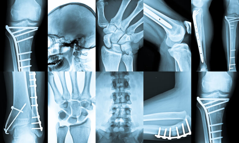
Moreover, the technology’s application extends beyond dentistry. It is increasingly used in ENT (Ear, Nose, and Throat) and orthopaedics for its capability to detail bone and joint structures. Its lower cost and accessibility make it an attractive option for smaller medical facilities or practices, thereby broadening the scope of advanced medical imaging available to a larger population.
Overall, CBCT technology represents a significant advancement in the medical field. It offers detailed imagery with lower health risks and enhances the efficacy and safety of medical diagnostics and treatments.
home » Cone Beam CT


