Windows into the Brain: The Expanding Role of Medical Imaging in Neurological Disorders
 By
By
Discover how medical imaging in neurological diseases revolutionises diagnosis and management of conditions like Alzheimer’s and Parkinson’s.
Dynamic Contrast Enhanced Magnetic Resonance Imaging (DCE-MRI) has emerged as a pivotal technique in the area of diagnostic imaging, offering unparalleled insights into tissue vascularity and perfusion. This advanced imaging modality leverages the administration of contrast agents to enhance the visualisation of blood flow dynamics within tissues, proving especially beneficial in oncology, neurology, and cardiology.
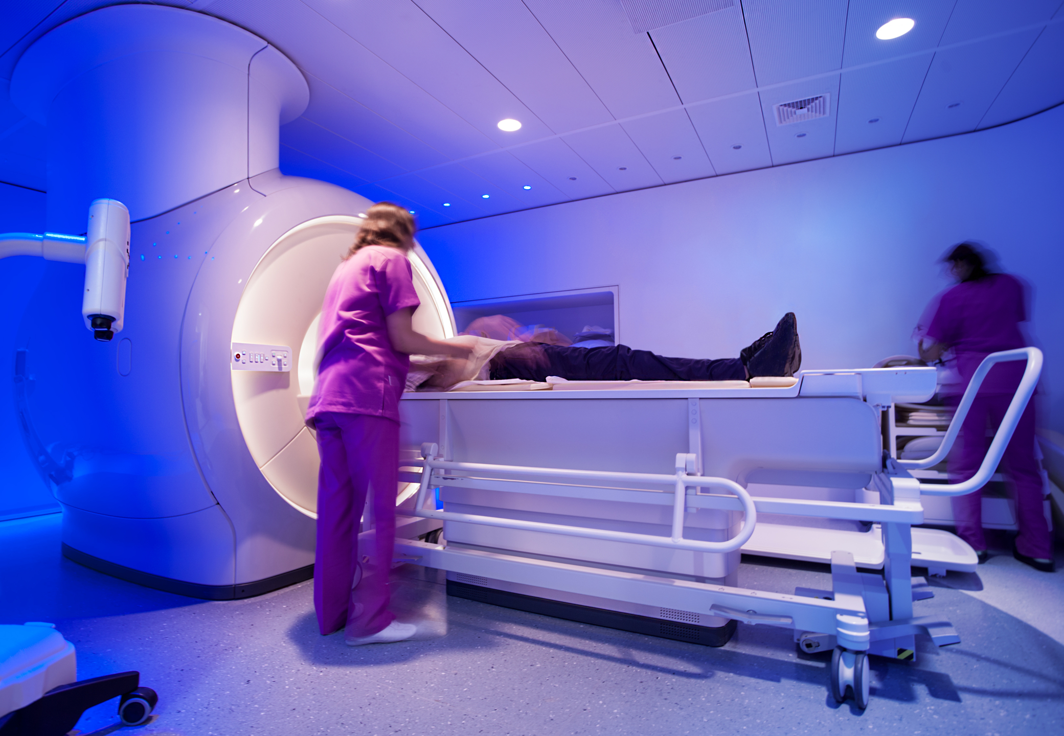
The core principle of DCE-MRI involves injecting a gadolinium-based contrast agent into the patient’s bloodstream. As the contrast agent circulates, it alters the magnetic properties of nearby water molecules, thereby enhancing the signal on MRI scans. The imaging process is conducted dynamically, capturing a series of images over time. This temporal aspect is crucial, as it allows radiologists to observe the rate at which the contrast agent is absorbed and washed out of tissues, providing vital information about tissue perfusion and capillary permeability.
In oncology, DCE-MRI has revolutionised the evaluation of tumours. Malignant tissues typically exhibit aberrant vascular properties, such as increased permeability and abnormal angiogenesis. By analysing the contrast enhancement patterns, radiologists can more accurately differentiate between benign and malignant lesions. Furthermore, DCE-MRI is instrumental in monitoring the efficacy of cancer therapies. Changes in tumour vascularity following treatment can be detected early, enabling prompt adjustments to therapeutic strategies.
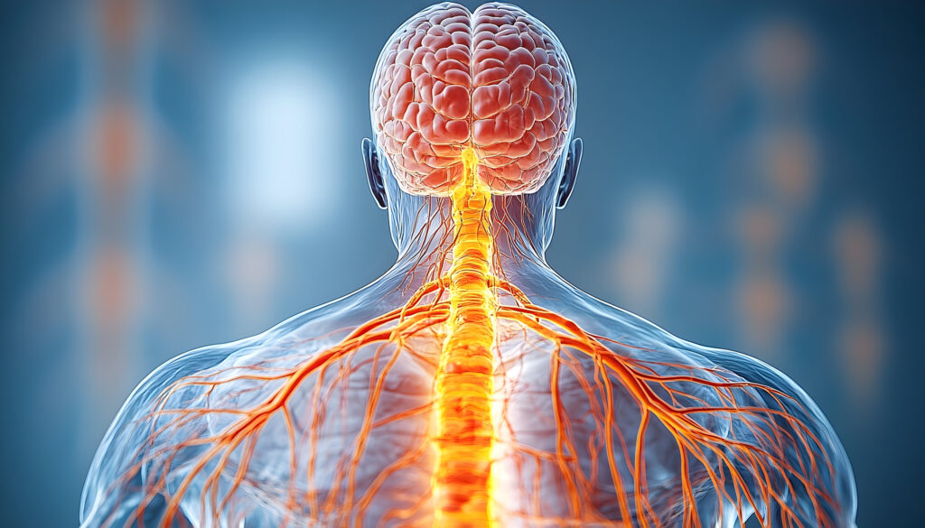
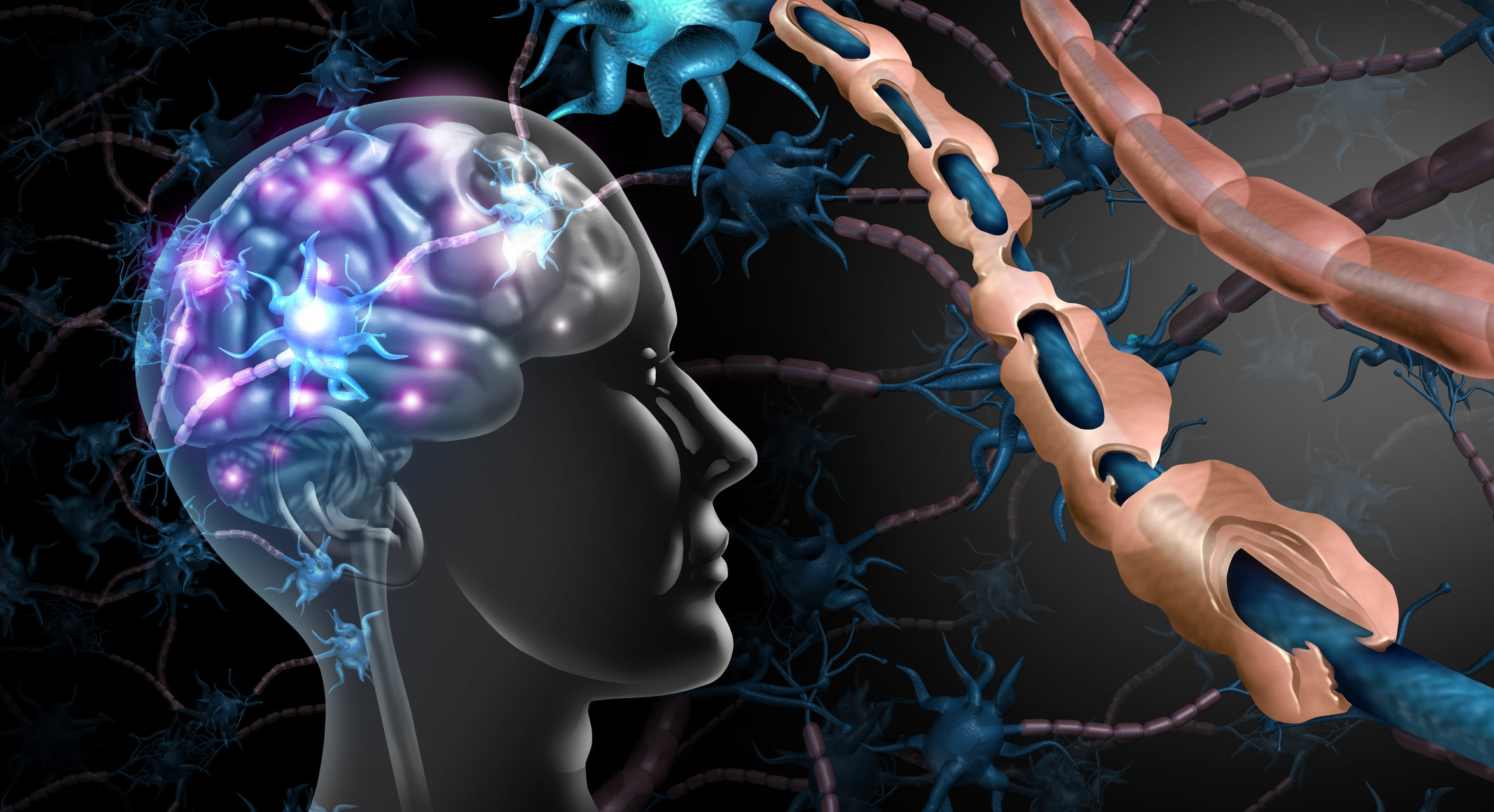
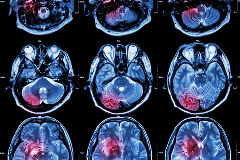
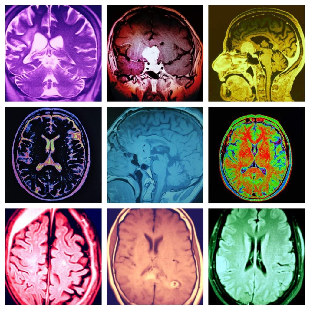
Neurological applications of DCE-MRI are equally transformative. This technique is utilised to assess cerebral blood flow and blood-brain barrier integrity, which are critical in diagnosing and managing conditions like stroke, multiple sclerosis, and brain tumours. By providing detailed maps of perfusion and vascular leakage, DCE-MRI aids in identifying regions of the brain affected by pathological processes, thus guiding clinical decision-making.
In cardiology, DCE-MRI plays a significant role in evaluating myocardial perfusion. It helps identify areas of ischaemia and infarction, offering a noninvasive means to assess the severity and extent of cardiac conditions. This information is vital for planning appropriate interventions and managing patient outcomes.
Even though it has numerous advantages, DCE-MRI is not without challenges. The need for contrast agents poses a risk for patients with renal impairment, and the interpretation of dynamic images requires advanced expertise. Nonetheless, ongoing advancements in imaging technology and analytical techniques continue to enhance the accuracy and safety of DCE-MRI.
Therefore, Dynamic Contrast Enhanced MRI stands as a cornerstone of modern diagnostic imaging. Its ability to provide detailed and dynamic insights into tissue vascularity and perfusion makes it an invaluable tool in diagnosing and managing a wide array of medical conditions.
home »
By
Discover how medical imaging in neurological diseases revolutionises diagnosis and management of conditions like Alzheimer’s and Parkinson’s.
 By
By
Explore the latest innovations in MRI equipment and how they enhance diagnostic imaging and improve patient care outcomes.
 By
By
Learn about medical imaging in multiple sclerosis: essential techniques for diagnosing, tracking, and managing the disease.
 By
By
Medical image quality plays a crucial role in ensuring accurate diagnoses, effective treatment plans, and improved patient care across healthcare systems. Image for illustration only. Person depicted is a model.
 By
By
Magnetic resonance imaging (MRI) revolutionized medical diagnostics by evolving from nuclear magnetic resonance discoveries to life-saving technology.
 By
By
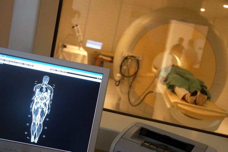
MRI scans provide critical insights into the body’s internal structures, aiding in the diagnosis and treatment of various conditions.
 By
By
Understanding the different types of MRI involves exploring specialised techniques like fMRI, dMRI, MRS, and CMR, each offering unique diagnostic insights.