Bridging the Gap Between Medical Imaging and Neurological Care
 By
By
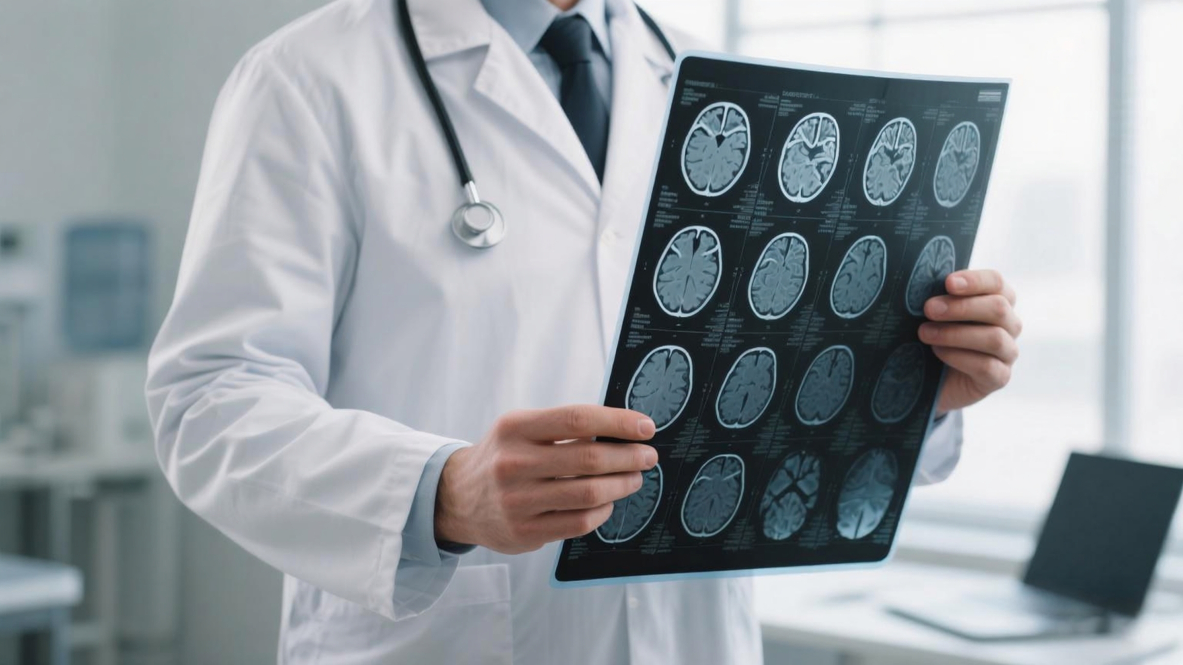
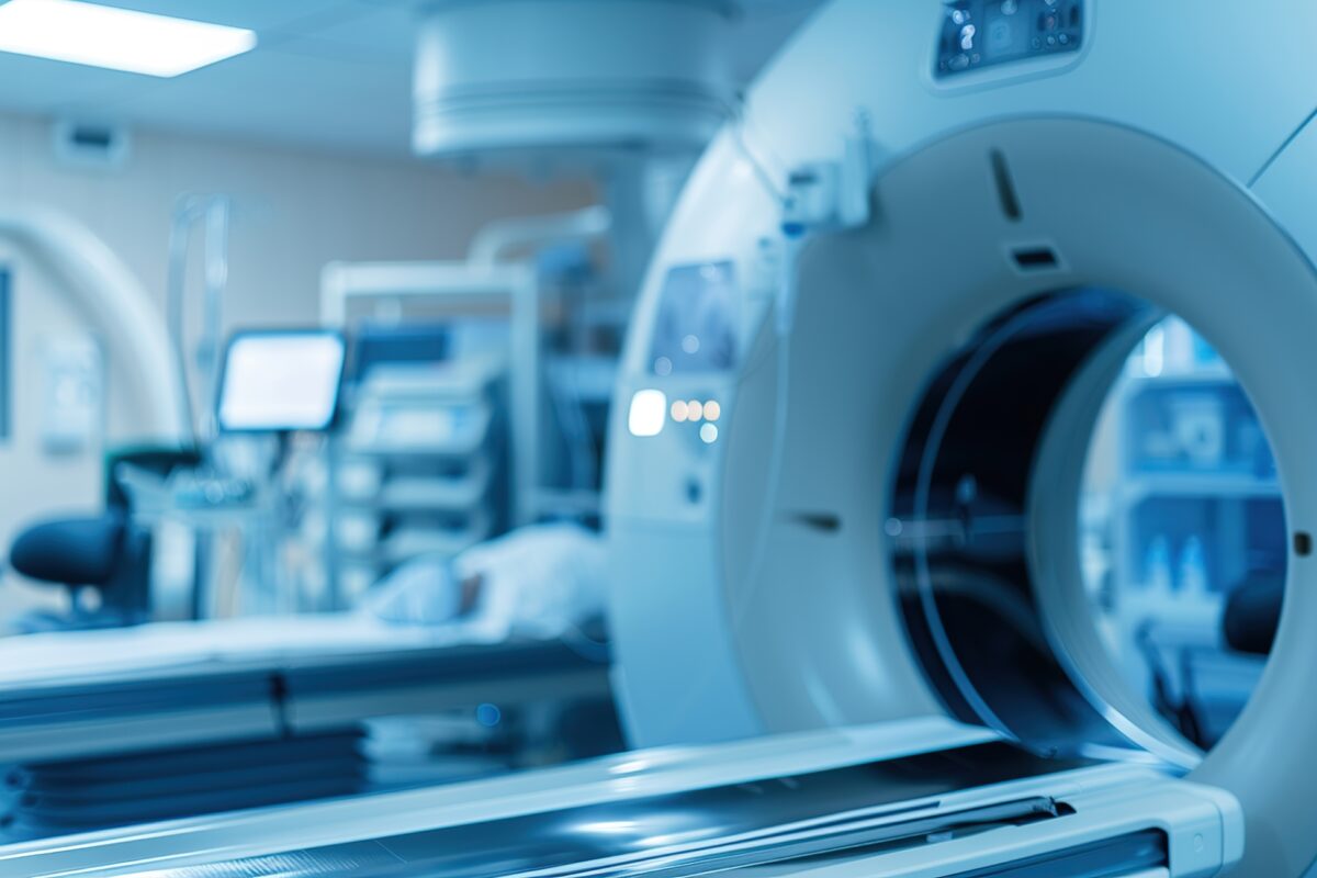
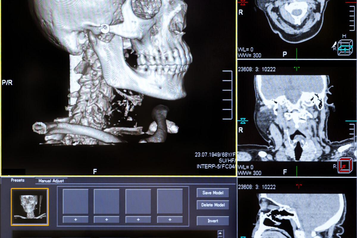
Discover the complexities of neurological care and the necessity for seamless information transfer in clinical practice.
By
Discover the complexities of neurological care and the necessity for seamless information transfer in clinical practice.
 By
By
Learn about Modern Medical Imaging and Radiation Therapy and how advanced technology shapes the future of oncological treatment.
 By
By
Navigate the complexities of medical treatment cost in Germany. Find out what influences pricing for different healthcare procedures.
 By
By
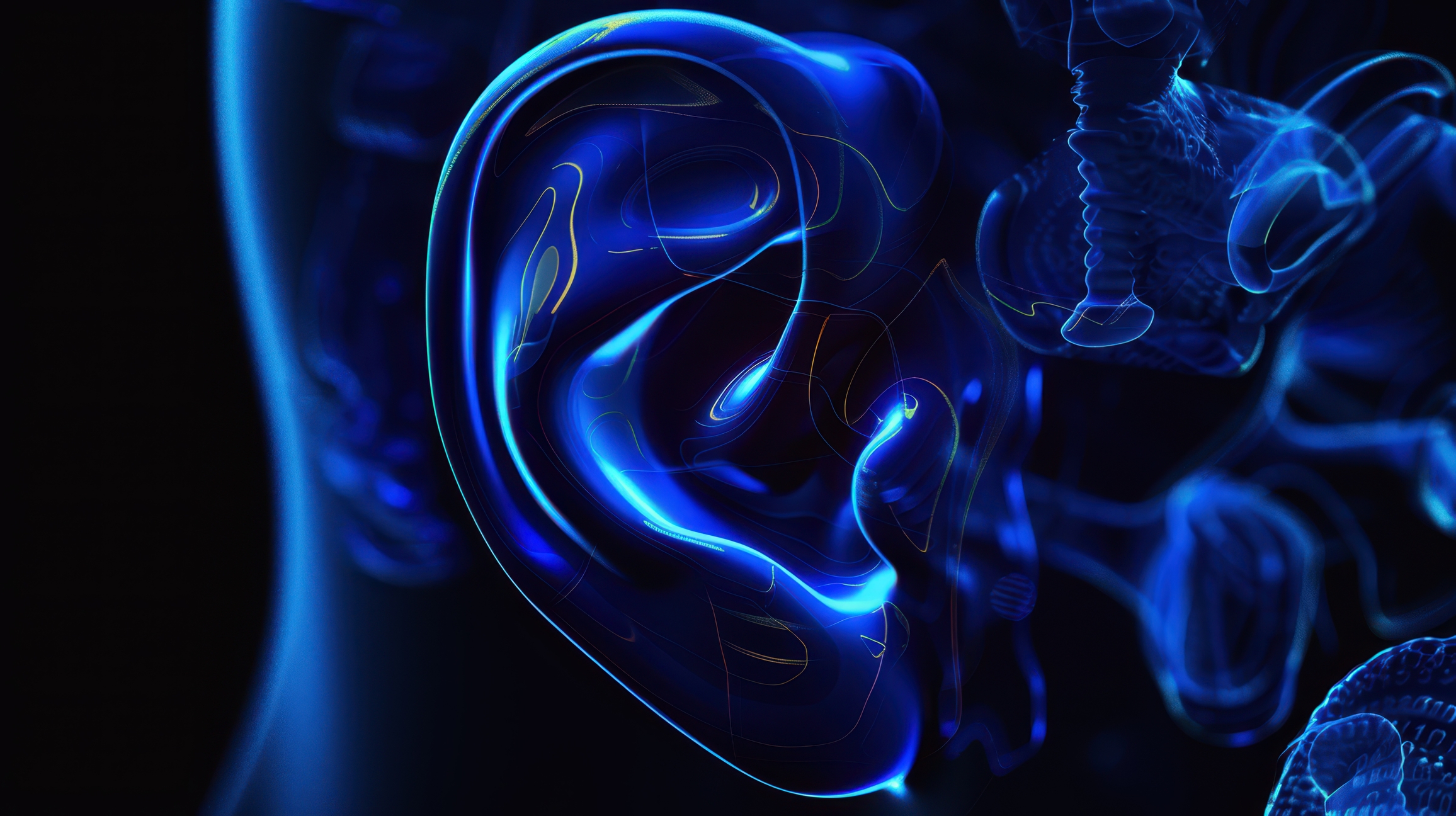
Discover how medical imaging of the ear transforms the evaluation of ear diseases and advances treatment options.
 By
By
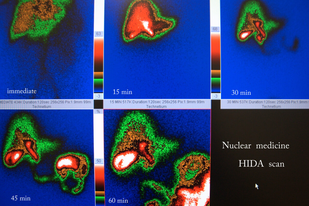
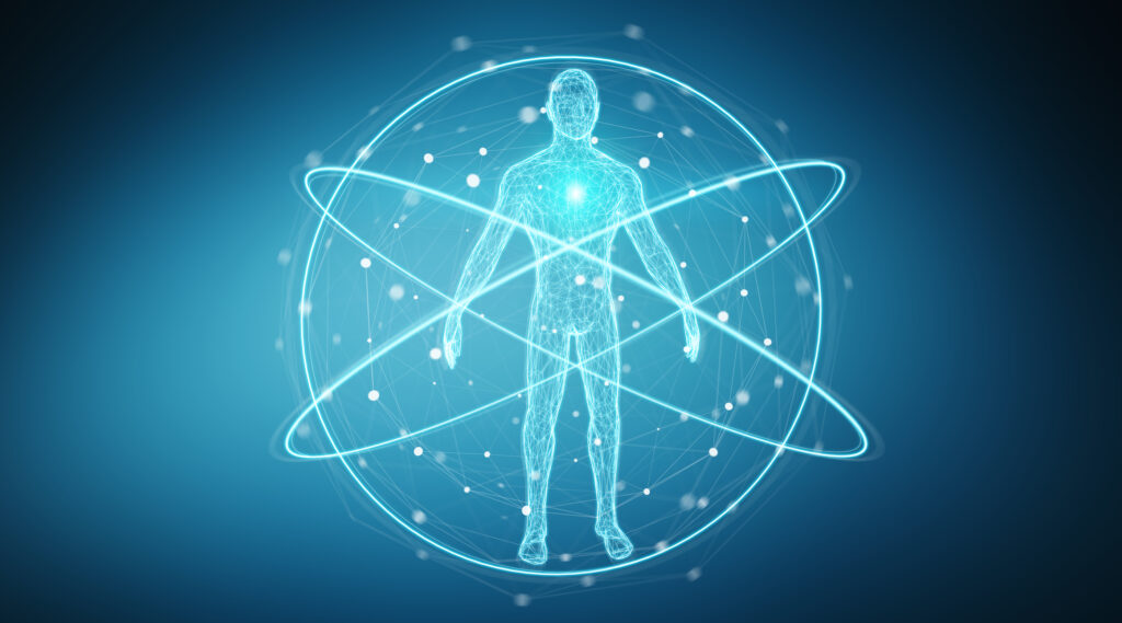
Discover how radiopharmaceuticals in medicine are transforming diagnostics and treatments with atomic precision for better health outcomes.
 By
By
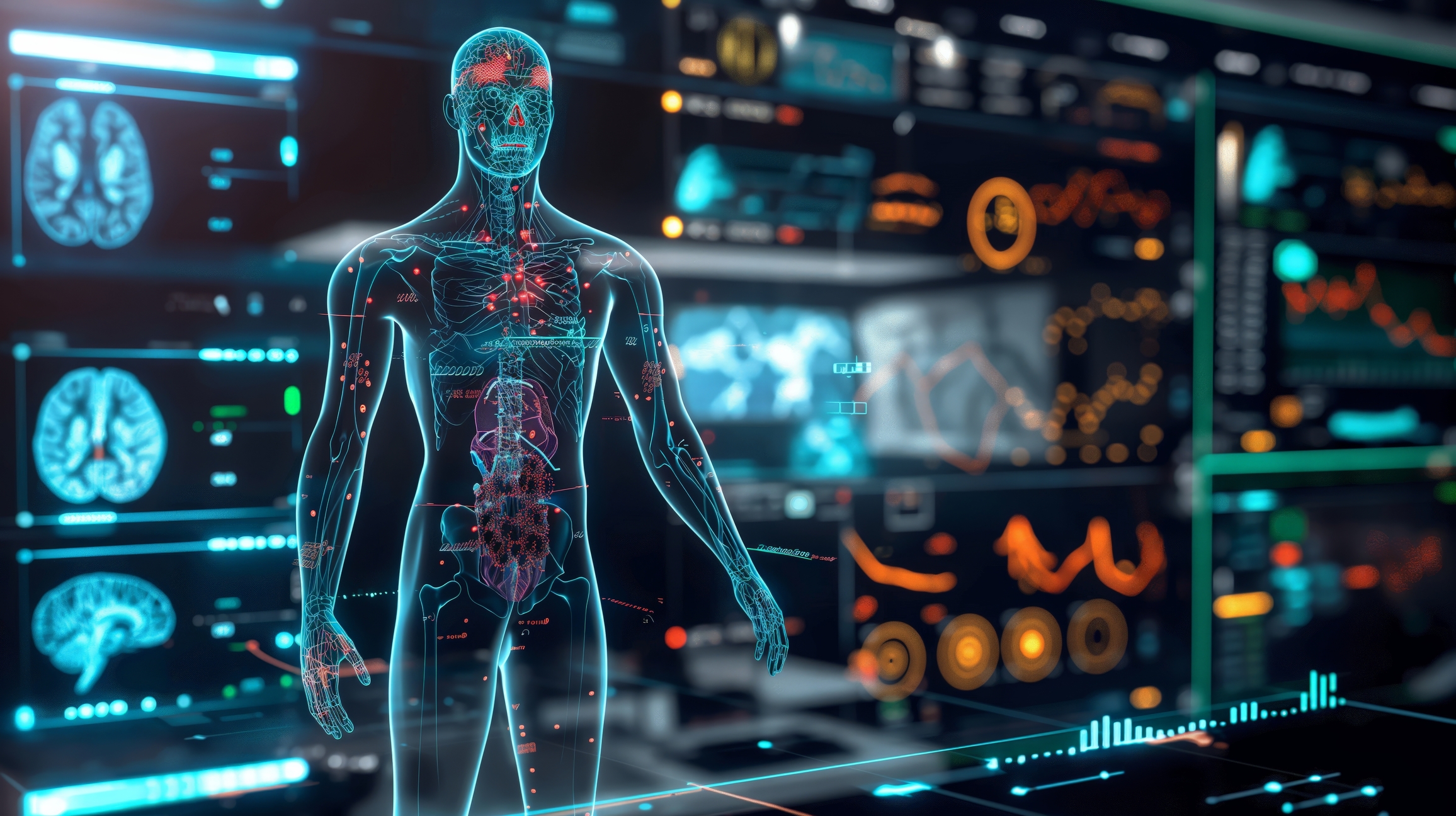
Discover the fascinating types of medical imaging, from X-rays to advanced molecular techniques, shaping health care today.
 By
By
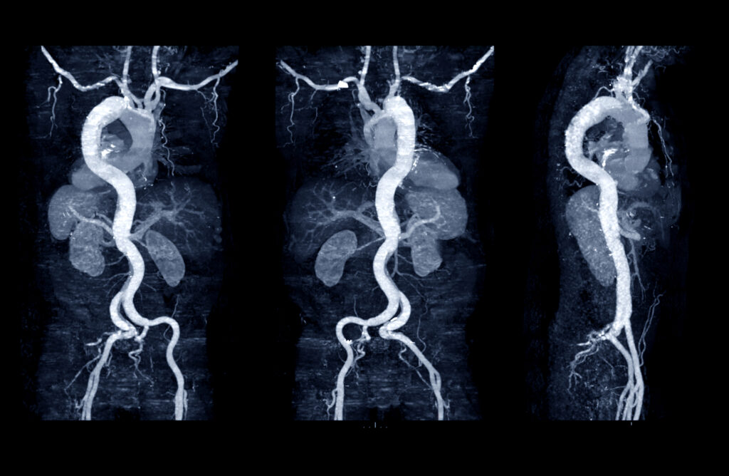
Discover the significance of the Aneurysm Progression Marker in predicting AAA growth with non-invasive perivascular fat density measurements.
 By
By
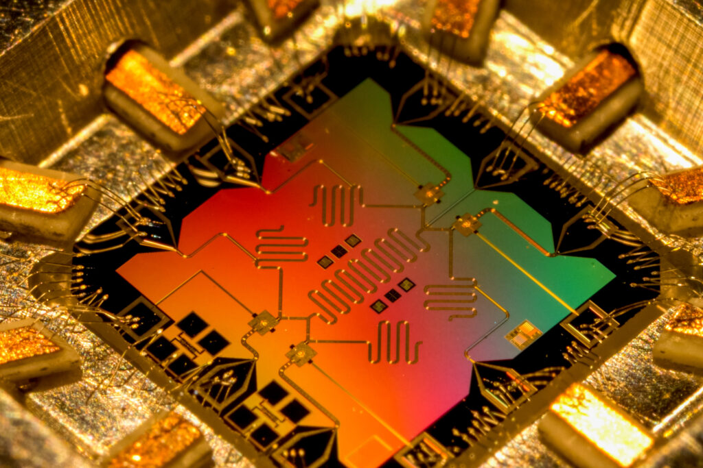
Find out how quantum computing in medical imaging is transforming diagnostics with enhanced algorithms and AI integration.
 By
By
Evaluate your expertise with the medical imaging quiz, focusing on imaging parameters, radiation safety, and practical case studies.
 By
By
Understand the strengths and limitations of Congenital neuroblastoma PET in assessing rare childhood tumors in this critical study.
 By
By
Understand the roles of medical scanners in modern medicine, highlighting their strengths and applications in healthcare diagnostics.
 By
By
Uncover the key uses of Carbon-14 radiotracers. Learn how they contribute to environmental science and biochemical studies.
 By
By
Learn about the exciting opportunities a career in radiochemistry offers in nuclear medicine and PET imaging. Image for illustration only. Person depicted is a model.
 By
By
Understand the role of regulation in nuclear medicine and its impact on patient safety and public confidence in healthcare.
 By
By
Learn about the UK Biobank’s pioneering imaging study that has delivered unique insights from 100,000 participant scans.
 By
By
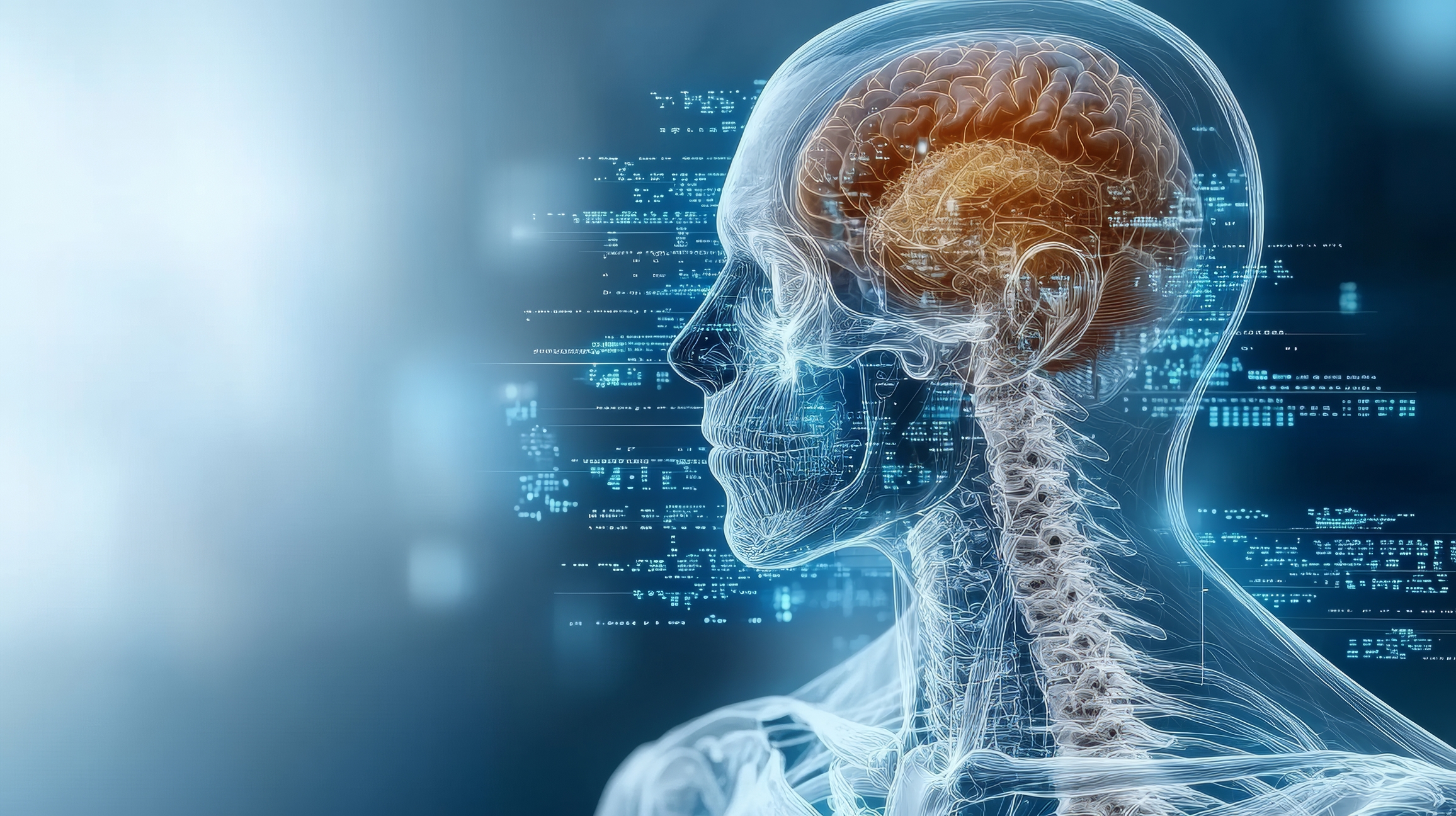
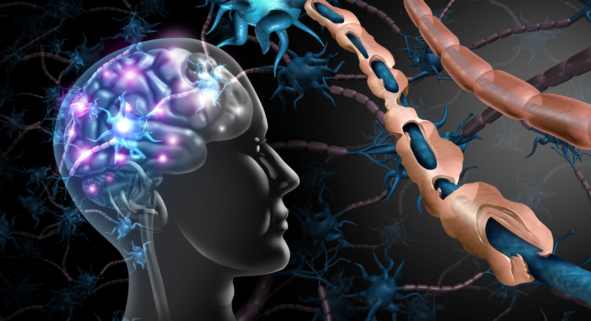
Neuroimaging techniques in drug side effects reveal how medications alter brain function, structure, and chemistry over time.
 By
By
Uncover the vital role of Diagnostic Imaging Physics in modern medicine with insights into imaging technologies and patient safety.
 By
By
Diagnostic imaging anxiety is a significant concern. Discover how it can influence mental health and healthcare outcomes. Image for illustration only. People depicted are models.
 By
By
Learn the impact of Imaging in Addiction Therapy, addressing challenges and offering insights into effective rehabilitation methods.
 By
By
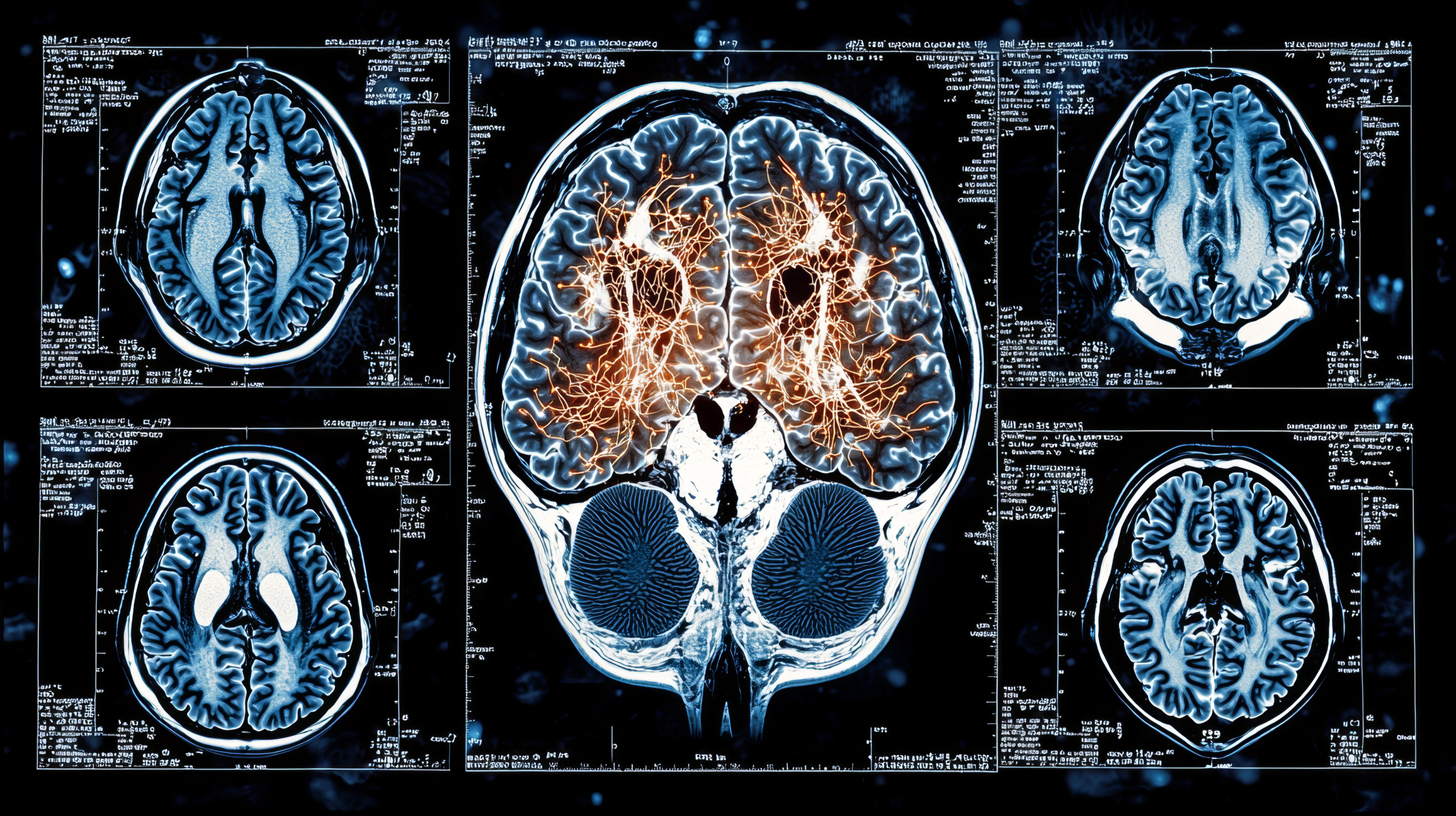
Learn about medical imaging in multiple sclerosis: essential techniques for diagnosing, tracking, and managing the disease.
 By
By
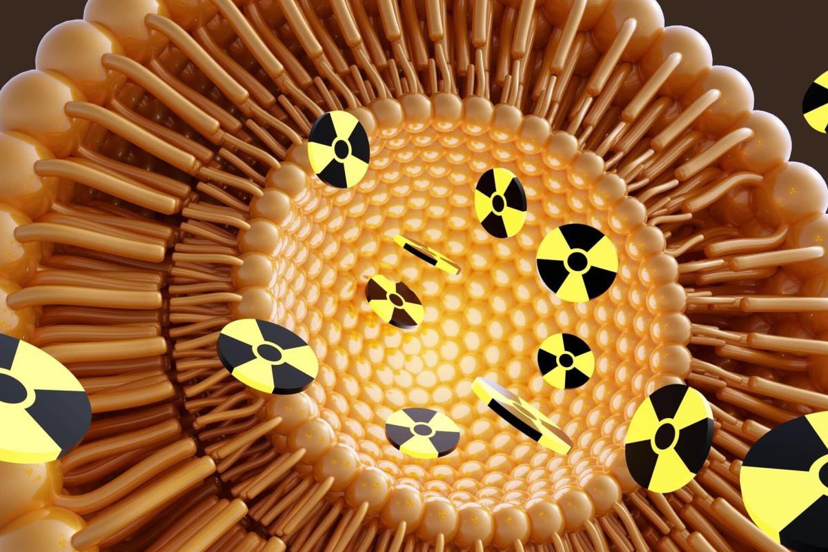
Radioactive isotopes emit targeted radiation that effectively destroys cancer cells while minimising harm to healthy tissues.
 By
By
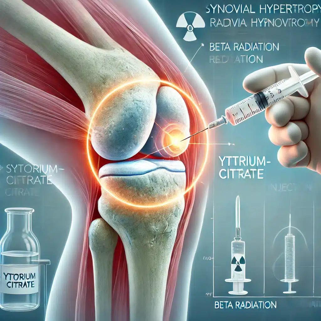
Yttrium-90 Citrate delivers targeted beta radiation, effectively treating synovial hypertrophy while minimising extra-articular radioactive escape risks.
 By
By
Zirconium-89 molecular imaging revolutionises diagnostics by enabling precise tumour localisation and tracking antibody-based therapies effectively.
 By
By
New Year medical imaging breakthroughs highlight innovative technologies, transforming healthcare diagnostics and improving patient outcomes worldwide.
 By
By
Medical imaging combines advanced technology, skilled professionals, and holiday warmth to support patients during Christmas emergencies.
 By
By
Sustainability in medical imaging highlights innovative practices, stakeholder roles, and strategies to reduce environmental impact effectively.
 By
By
Open MedScience fosters global collaboration, accelerates innovation, promotes transparency, enhances accessibility, and transforms healthcare outcomes.
 By
By
AI algorithms revolutionise tumour detection in medical imaging, enhancing precision, automating analysis, and supporting personalised cancer treatment through advanced PET/CT integration.
 By
By
Radionuclide Therapy Effects include potential organ toxicity, requiring careful monitoring to manage patient outcomes effectively.
 By
By
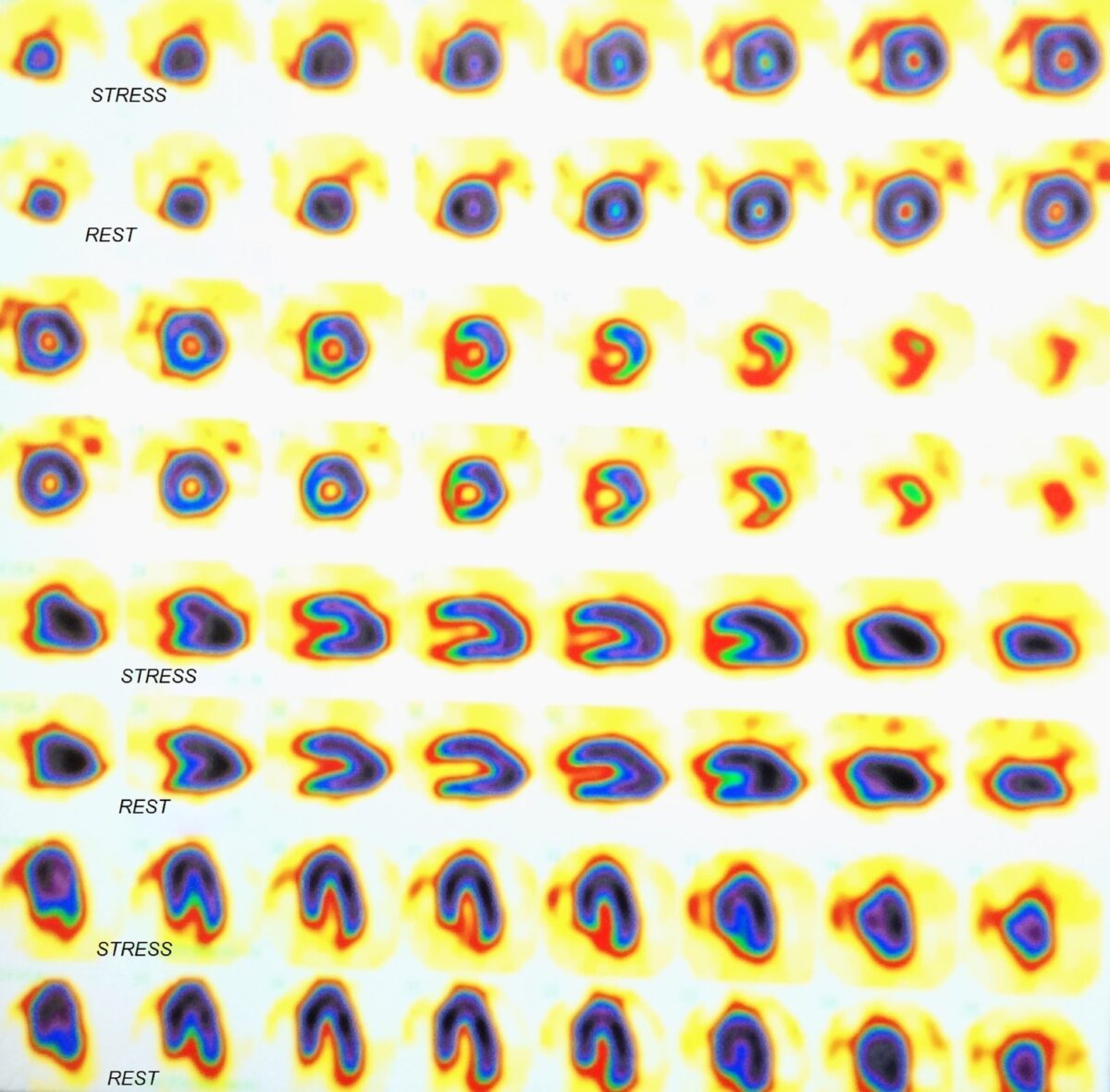
FLYRCADO, a radiopharmaceutical agent employed in PET myocardial perfusion imaging, provides accurate quantification of myocardial blood flow, enhancing diagnostic confidence.