Gamma Camera
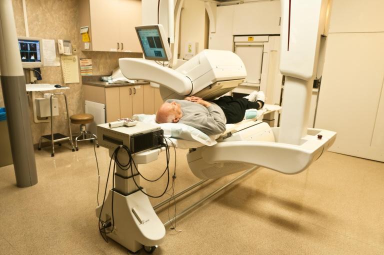
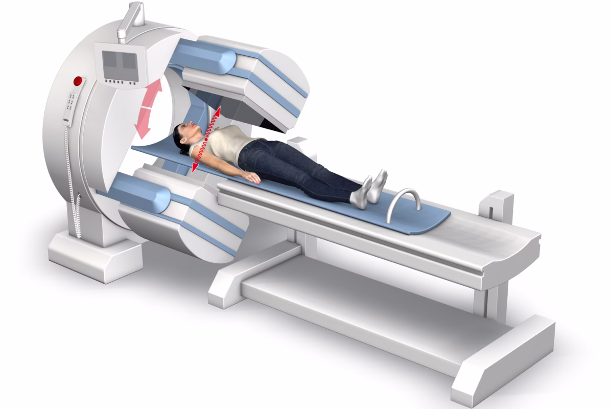
The gamma camera, an essential instrument in nuclear medicine, plays a pivotal role in diagnosing various diseases, including cancer and heart conditions. This sophisticated device captures gamma rays emitted from a patient’s body after the administration of a radioactive tracer, facilitating the creation of detailed images of organs and tissues.
Developed in the 1950s by Hal Anger, the gamma camera has undergone significant advancements over the decades. Its primary detector component includes a large, flat crystal that absorbs the gamma rays, converting them into light. This light is then amplified by photomultiplier tubes to produce a detailed image that specialists can analyse.
The gamma camera operates based on the principle of scintigraphy. The process involves the injection of a radiopharmaceutical into the patient’s bloodstream, which then travels to specific organs. The camera captures the emitted gamma rays, creating an image that highlights areas of high radiotracer concentration. These areas may indicate abnormal organ function or the presence of disease, providing crucial information for diagnosis and treatment planning.
One of the significant advantages of using a gamma camera in medical diagnostics is its ability to monitor physiological functions in real-time, offering insights that are often unattainable through other imaging techniques. Furthermore, the camera can perform scans over large areas of the body, providing comprehensive data with minimal discomfort to the patient.
As medical technology continues to evolve, the gamma camera remains a cornerstone in the field of nuclear medicine, aiding in the effective diagnosis and management of various health conditions. Its ability to provide precise and functional imaging ensures that it will continue to be a valuable asset in healthcare for years to come.
home » Gamma Camera
 By
By