Liver Fibrosis Imaging
Liver fibrosis is a progressive condition characterised by the excessive accumulation of extracellular matrix proteins, primarily collagen, in the liver. This condition can result from chronic liver diseases such as hepatitis B and C, alcohol-related liver disease, and non-alcoholic fatty liver disease (NAFLD). If left unchecked, fibrosis can advance to cirrhosis and liver failure, necessitating early detection and monitoring to prevent irreversible damage. Imaging techniques play a vital role in the non-invasive assessment of liver fibrosis, offering an alternative to the traditional gold standard of liver biopsy.
Non-Invasive Imaging Modalities
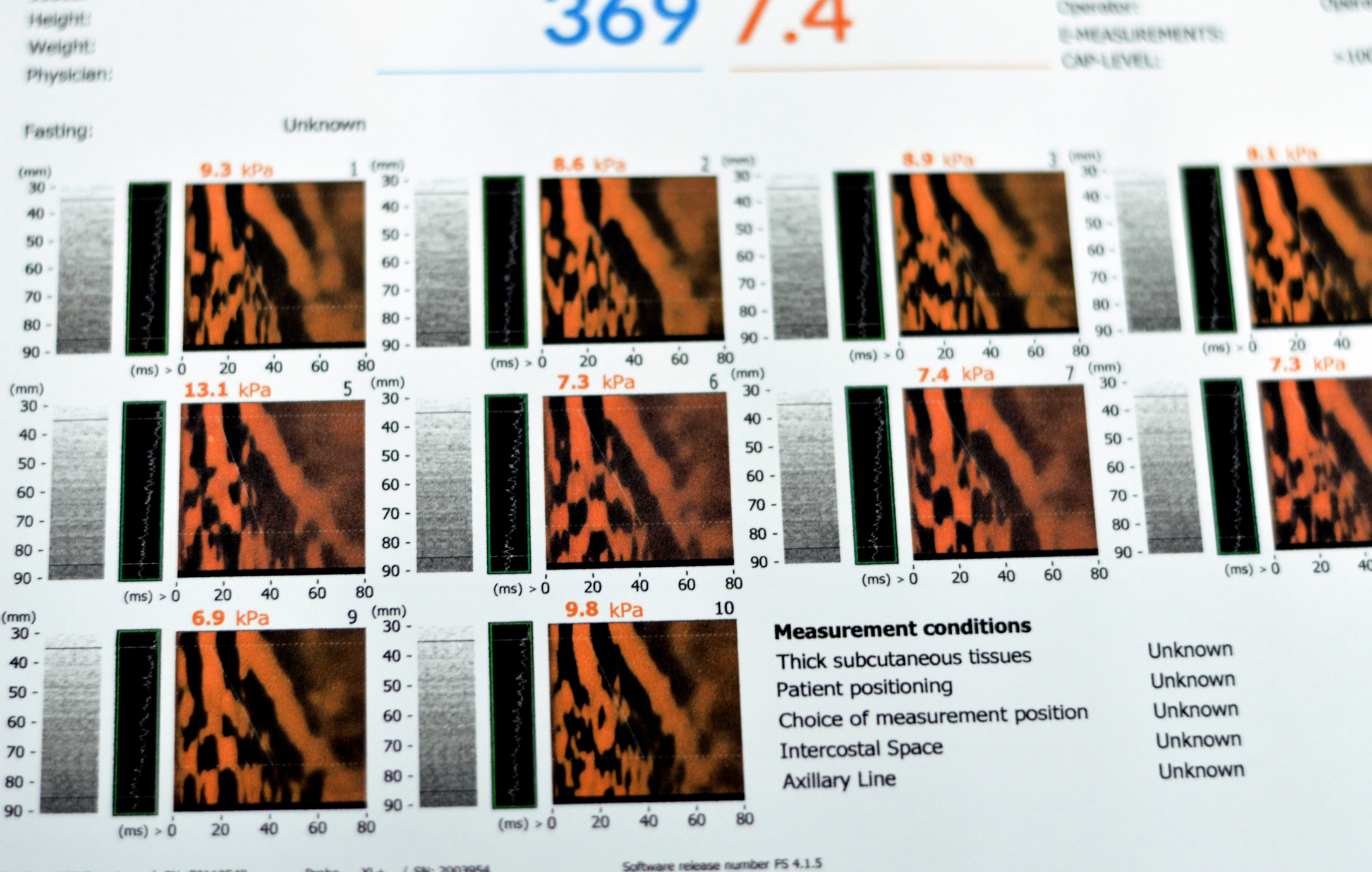
Non-invasive imaging techniques have revolutionised the diagnosis and management of liver fibrosis, minimising the risks associated with biopsy. Ultrasound-based elastography, including transient elastography (TE) and shear wave elastography (SWE), has become a cornerstone in clinical practice. These methods measure liver stiffness, which correlates with the degree of fibrosis. TE, commonly performed using devices like FibroScan, is widely used due to its simplicity and reproducibility. However, it may be less effective in individuals with obesity or ascites.
Magnetic resonance elastography (MRE) is another cutting-edge technique that provides a more detailed and comprehensive assessment of liver stiffness. MRE uses low-frequency mechanical waves in conjunction with MRI imaging, allowing for a highly accurate evaluation of fibrosis stages. It is particularly beneficial for patients with complex conditions or co-morbidities that limit the utility of ultrasound.
Advanced Imaging Techniques
Beyond elastography, other imaging modalities contribute to the diagnosis and monitoring of liver fibrosis. Contrast-enhanced ultrasound (CEUS) and computed tomography (CT) can evaluate liver perfusion changes associated with advanced fibrosis and cirrhosis. However, these techniques are less specific for early-stage fibrosis and are generally not first-line choices.
Magnetic resonance imaging (MRI) techniques, including T1 and T2-weighted imaging and diffusion-weighted imaging (DWI), are also utilised in liver fibrosis assessment. These methods provide valuable information on tissue characteristics and inflammation. Moreover, the advent of hepatocyte-specific contrast agents and quantitative imaging biomarkers has improved the ability to detect fibrosis at earlier stages.
Emerging Technologies and Applications
Artificial intelligence (AI) and machine learning algorithms are being integrated into imaging workflows, enhancing diagnostic accuracy and enabling automated fibrosis quantification. These technologies can analyse complex imaging data, identify subtle patterns, and support clinical decision-making.
Conclusion
Liver fibrosis imaging has evolved significantly, providing non-invasive, reliable, and patient-friendly options for diagnosis and monitoring. As technology advances, imaging modalities such as MRE and AI-driven tools are expected to play an increasingly prominent role in personalised treatment strategies. These innovations not only reduce the reliance on invasive procedures but also empower clinicians to optimise care for patients with chronic liver diseases.
home » Liver Fibrosis Imaging

 By
By