Medical Imaging in Therapy Planning
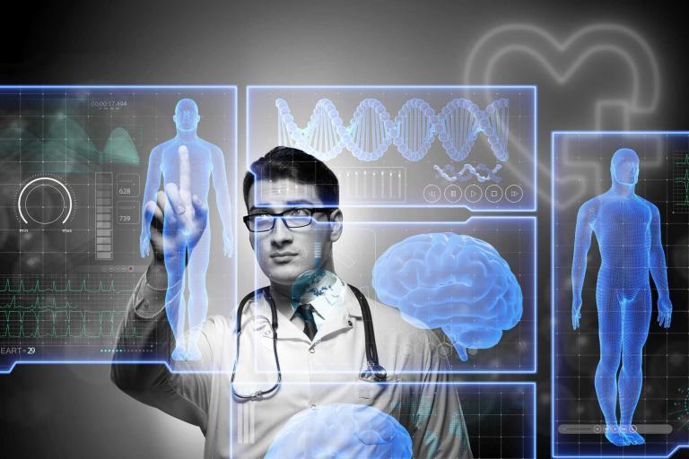
Medical imaging plays a pivotal role in therapy planning across various medical disciplines, significantly enhancing the precision and effectiveness of treatment protocols. By providing detailed visualisation of anatomical structures and physiological processes, medical imaging allows clinicians to develop tailored therapy plans that address each patient’s unique needs.
One of the foremost applications of medical imaging in therapy planning is in oncology. Imaging modalities such as computed tomography (CT), magnetic resonance imaging (MRI), and positron emission tomography (PET) are indispensable tools in the diagnosis, staging, and monitoring of cancer. These technologies enable the accurate delineation of tumours, helping in the precise targeting of radiotherapy. For instance, CT and MRI scans provide high-resolution images of tumour boundaries and surrounding tissues, allowing oncologists to design radiation fields that maximise tumour destruction while sparing healthy tissue. PET scans, on the other hand, offer functional imaging that can identify the metabolic activity of cancer cells, further refining the targeting process.
In addition to oncology, medical imaging is crucial in cardiovascular therapy planning. Techniques like echocardiography, cardiac MRI, and coronary angiography provide detailed images of the heart and blood vessels, facilitating the diagnosis and treatment of cardiovascular diseases. For example, echocardiography uses ultrasound waves to create real-time images of heart function, aiding in the assessment of heart valve disorders and cardiomyopathies. Cardiac MRI offers comprehensive data on myocardial structure and function, which is vital for planning interventions such as angioplasty or bypass surgery.
Neurosurgery also benefits immensely from advanced imaging techniques. Preoperative planning for brain and spinal surgeries relies heavily on MRI and CT scans. These modalities provide high-resolution images of neural structures, allowing neurosurgeons to map out surgical approaches with utmost precision. Functional MRI (fMRI) and diffusion tensor imaging (DTI) further enhance surgical planning by mapping brain activity and neural pathways, thereby minimising the risk of damage to critical areas during surgery.
Furthermore, medical imaging aids in the management of musculoskeletal conditions. MRI and ultrasound are particularly valuable in diagnosing and planning treatment for injuries and disorders affecting muscles, joints, and bones. MRI provides detailed images of soft tissues, making it an excellent tool for assessing ligament tears, cartilage damage, and other soft tissue injuries. With its real-time imaging capability, ultrasound is often used for guiding injections and other minimally invasive procedures.
In summary, medical imaging is an integral component of therapy planning, offering invaluable insights into the structure and function of the human body. Its ability to provide detailed and accurate images ensures that treatment plans are precisely tailored to each patient’s condition, ultimately improving clinical outcomes and enhancing the quality of care. As imaging technologies continue to evolve, their role in therapy planning is expected to become even more significant, paving the way for more personalised and effective medical treatments.
home » Medical Imaging in Therapy Planning

 By
By