Prostate Fusion Biopsy Imaging
Prostate fusion biopsy imaging is a modern technique used to enhance the accuracy of prostate cancer diagnosis. It combines multiparametric magnetic resonance imaging (mpMRI) with real-time ultrasound (US) to create a highly detailed and precise image of the prostate, allowing targeted biopsy sampling. This method represents a significant advancement over traditional systematic biopsy approaches, which rely solely on transrectal ultrasound (TRUS) and often result in missed cancers or unnecessary biopsies.
The Need for Fusion Biopsy
Prostate cancer is one of the most common malignancies in men, and early detection is crucial for effective treatment. Conventional TRUS-guided biopsies can be unreliable, as they involve taking multiple random samples from different areas of the prostate. This random approach can lead to underdiagnosis, particularly in patients with small or anteriorly located tumours, or overdiagnosis of clinically insignificant cancers. The advent of mpMRI allows for better visualisation of suspicious lesions, but MRI alone is not sufficient for biopsy. Fusion biopsy bridges this gap by enabling the precise targeting of abnormal regions seen on MRI.
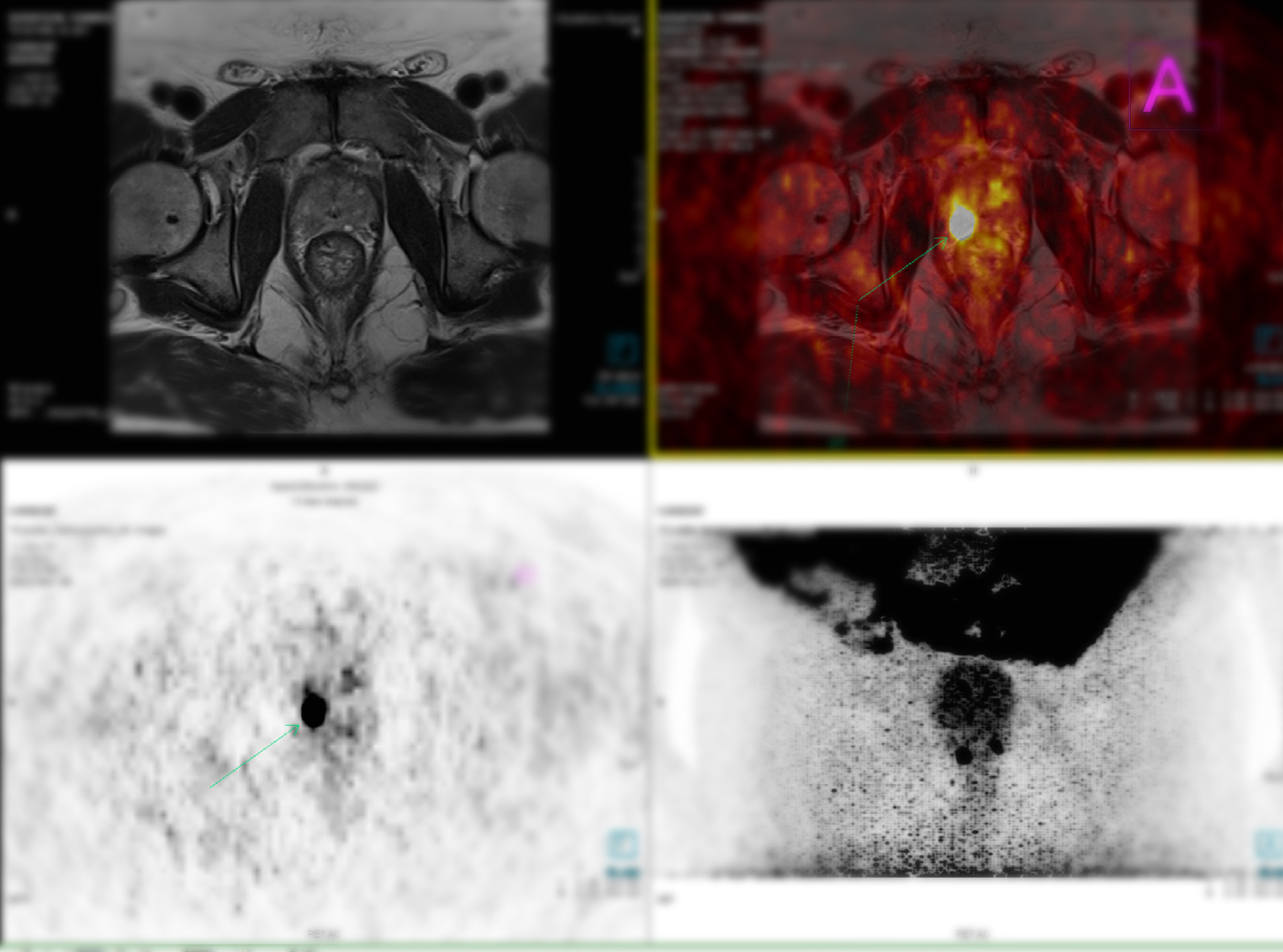
How Prostate Fusion Biopsy Works
The process begins with an mpMRI scan, which provides high-resolution images of the prostate and identifies regions of interest (ROI) based on the Prostate Imaging-Reporting and Data System (PI-RADS) scoring system. These images are then fused with real-time ultrasound during the biopsy procedure. There are two primary methods of performing a prostate fusion biopsy:
- Cognitive Fusion: The radiologist visually correlates the MRI images with the ultrasound images, guiding the biopsy based on their expertise.
- Software-Assisted Fusion: Advanced software overlays the MRI images onto the real-time ultrasound images, providing a more precise, computer-assisted guide for the biopsy.
The biopsy can be performed via either the transrectal or transperineal approach. The transperineal route is increasingly preferred due to its lower infection risk and ability to access anterior prostate lesions more effectively.
Advantages of Fusion Biopsy
Fusion biopsy offers several advantages over conventional TRUS-guided biopsy:
- Higher Accuracy: It increases the detection rate of clinically significant prostate cancer while reducing the likelihood of missing aggressive tumours.
- Reduced Unnecessary Biopsies: Patients with low-risk lesions on MRI may avoid unnecessary biopsies, reducing complications such as bleeding, infection, and discomfort.
- Lower Infection Risk: The transperineal approach used in fusion biopsy lowers the risk of sepsis compared to traditional transrectal biopsies.
- Personalised Patient Management: Fusion biopsy results help guide treatment decisions, distinguishing between patients who require active surveillance and those needing definitive therapy.
Conclusion
Prostate fusion biopsy imaging represents a major step forward in prostate cancer diagnostics. By integrating MRI and ultrasound, it enhances the precision and reliability of prostate biopsies, improving patient outcomes while minimising risks. As technology advances, further refinements in fusion imaging and artificial intelligence-assisted interpretation may continue to improve detection and management strategies.
home » Prostate Fusion Biopsy Imaging

 By
By