MRI Equipment Breakthroughs: How 2025 Innovations Transform Patient Care
 By
By
Explore the latest innovations in MRI equipment and how they enhance diagnostic imaging and improve patient care outcomes.
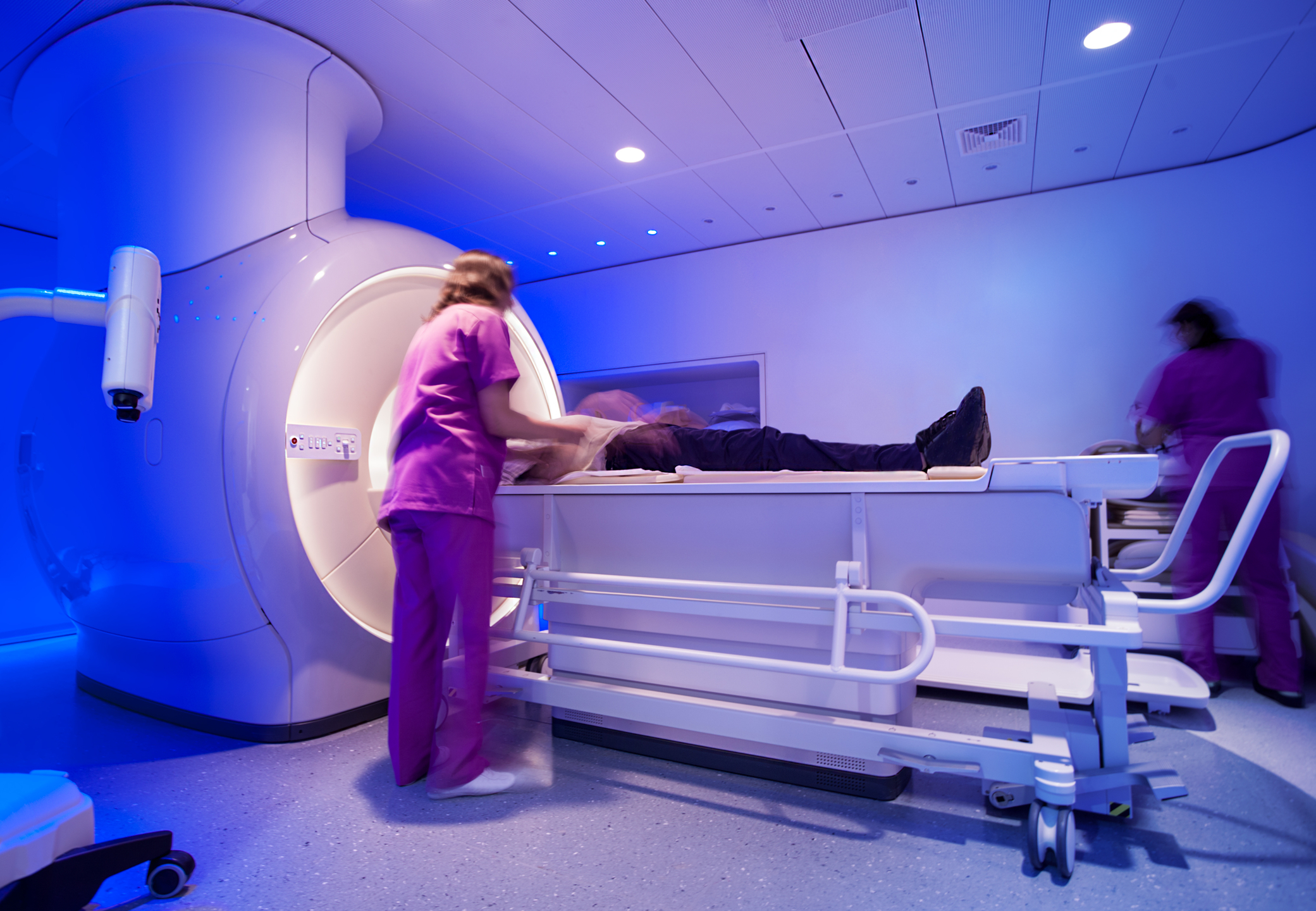
Real-Time Functional MRI (rt-fMRI) is an advanced neuroimaging technique that allows the immediate analysis and visualisation of brain activity as it occurs. Unlike conventional fMRI, which is typically used for post-hoc analysis of brain function, rt-fMRI provides dynamic feedback, enabling a range of applications in neuroscience, clinical medicine, and brain-computer interfaces.
Principles of Real-Time Functional MRI
Like standard fMRI, real-time fMRI relies on the blood-oxygen-level-dependent (BOLD) signal, which reflects changes in neural activity. When a brain region becomes active, the local demand for oxygen increases, leading to a measurable shift in the ratio of oxyhaemoglobin to deoxyhaemoglobin. Traditional fMRI studies collect large datasets over a period of time, with data processing and analysis occurring after the scanning session. In contrast, rt-fMRI employs rapid data acquisition and online processing to provide near-instantaneous feedback, often within seconds of the brain activity occurring.
Real-time processing involves several key steps: image reconstruction, motion correction, signal filtering, and statistical analysis. Advanced computational algorithms, including machine learning approaches, are increasingly being integrated to enhance speed and accuracy. The processed data can then be displayed to participants in real time, making it possible to use brain activity as a direct input for behavioural or therapeutic interventions.
Applications of Real-Time fMRI
Neurofeedback Training
One of the most widely explored applications of rt-fMRI is neurofeedback training. In this approach, individuals are trained to regulate their own brain activity by receiving real-time visual or auditory feedback. For example, participants may be shown a dynamic representation of their brain activity and instructed to modulate it through mental strategies such as visualisation or focused attention. This technique has been investigated for conditions such as depression, anxiety disorders, chronic pain, and schizophrenia. Studies suggest that individuals can learn to exert voluntary control over specific brain regions, potentially leading to lasting therapeutic benefits.
Brain-Computer Interfaces (BCIs)
Real-time fMRI can be incorporated into brain-computer interfaces, which enable direct communication between the brain and external devices. While electroencephalography (EEG) is more commonly used in BCIs due to its high temporal resolution, rt-fMRI offers superior spatial resolution, making it useful for applications requiring precise localisation of brain activity.
Clinical and Research Applications
Rt-fMRI has significant potential in clinical neuroscience. It can be used to monitor brain activity during surgery, assess cognitive function in minimally conscious patients, or track the progression of neurological disorders. In research, it allows the study of dynamic brain processes, such as decision-making, learning, and emotional regulation, in a way that traditional fMRI cannot.
Challenges and Future Directions
Despite its potential, real-time fMRI faces technical and practical challenges. High computational demands, artefacts from motion, and limitations in temporal resolution are key issues. Improvements in MRI hardware, machine learning algorithms, and signal processing techniques are expected to enhance its utility. In the future, rt-fMRI may play an even greater role in personalised medicine, mental health interventions, and human-computer interaction.
home » Real-Time Functional MRI
Explore the latest innovations in MRI equipment and how they enhance diagnostic imaging and improve patient care outcomes.