Medical Imaging Techniques for the Optic Nerve
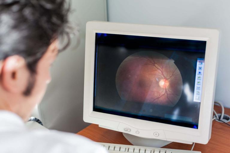
Medical imaging, crucial for optic nerve disorders, has evolved with technologies like MRI and OCT. Image for illustration only. Person depicted is a model.
Visual evoked potential (VEP) is a non-invasive diagnostic tool used to assess the visual pathway’s functional integrity objectively. The technique involves using specialized equipment to measure electrical activity in the brain in response to visual stimuli. VEP is beneficial for diagnosing and monitoring various ocular and neurological disorders, as well as aiding in evaluating visual acuity and determining the visual field.
The human visual system is a complex network of neural pathways responsible for processing and interpreting visual information. The visual path begins with the photoreceptors in the retina, which detect light and transmit the visual signal to the brain via the optic nerve. The signal then travels through various brain regions, ultimately reaching the primary visual cortex, where it is processed and interpreted.
Visual evoked potential testing involves the presentation of specific visual stimuli to the patient, usually in the form of checkerboard patterns, which alternate in contrast at varying frequencies. Electrodes on the scalp record the brain’s electrical activity in response to these stimuli, generating a waveform. This waveform is then analyzed to determine the amplitude and latency of the response, which provides valuable information about the functional integrity of the visual pathway.
One of the primary applications of VEP is in diagnosing and monitoring optic neuritis, an optic nerve inflammation often associated with multiple sclerosis (MS). VEP typically reveals prolonged latency and reduced amplitude in patients with optic neuritis, indicating slowed or impaired signal transmission along the visual pathway. VEP can also be used to differentiate between optic nerve disorders and other causes of visual loss, such as retinal disorders or macular degeneration.
Another important application of VEP is in evaluating visual acuity and determining the visual field. In patients with unexplained vision loss or poor visual acuity, VEP can provide objective evidence of the presence or absence of a visual pathway disorder. This information is particularly useful in pediatric populations, where traditional visual acuity testing may be challenging or unreliable.
In addition to its diagnostic applications, VEP is also used in various research settings to investigate the neurophysiological basis of visual perception and processing. Studies employing VEP have provided valuable insights into the neural mechanisms underlying various aspects of vision, such as contrast sensitivity, spatial frequency processing, and the effects of the ageing visual system.
Medical imaging, crucial for optic nerve disorders, has evolved with technologies like MRI and OCT. Image for illustration only. Person depicted is a model.
