Contact Lenses Used to Work Fine, So What Changed?
 By
By
Learn about contact lens intolerance and its symptoms. Discover what to do when lenses become uncomfortable during daily life.
By
Learn about contact lens intolerance and its symptoms. Discover what to do when lenses become uncomfortable during daily life.
 By
By
Flashes and floaters may signal retinal detachment. Discover a practical approach to imaging and timely surgical planning.
 By
By
Learn how indoor gross motor activities can help children develop vital physical skills while having fun in any living space.
 By
By
Get the most from your eye care visits. Learn key questions to ask eye care providers about diagnosis and treatment options.
 By
By
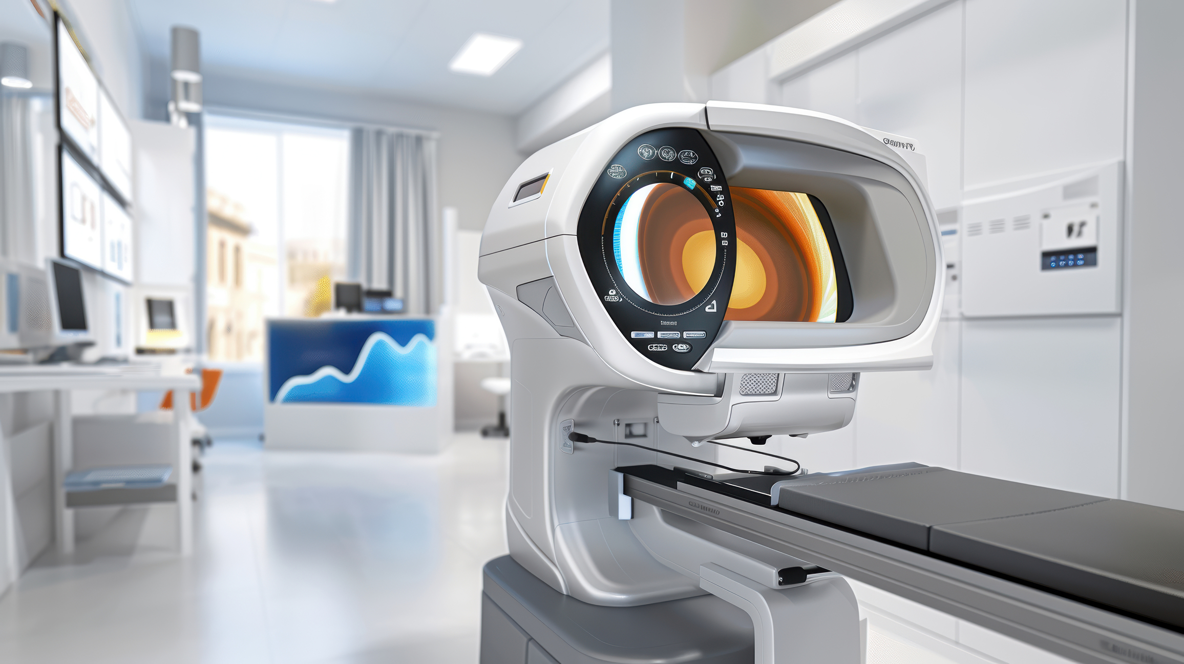
Discover the role of optical imaging in healthcare, supporting early disease detection and improved decision-making for clinicians.
 By
By
Discover the fascinating types of medical imaging, from X-rays to advanced molecular techniques, shaping health care today.
 By
By
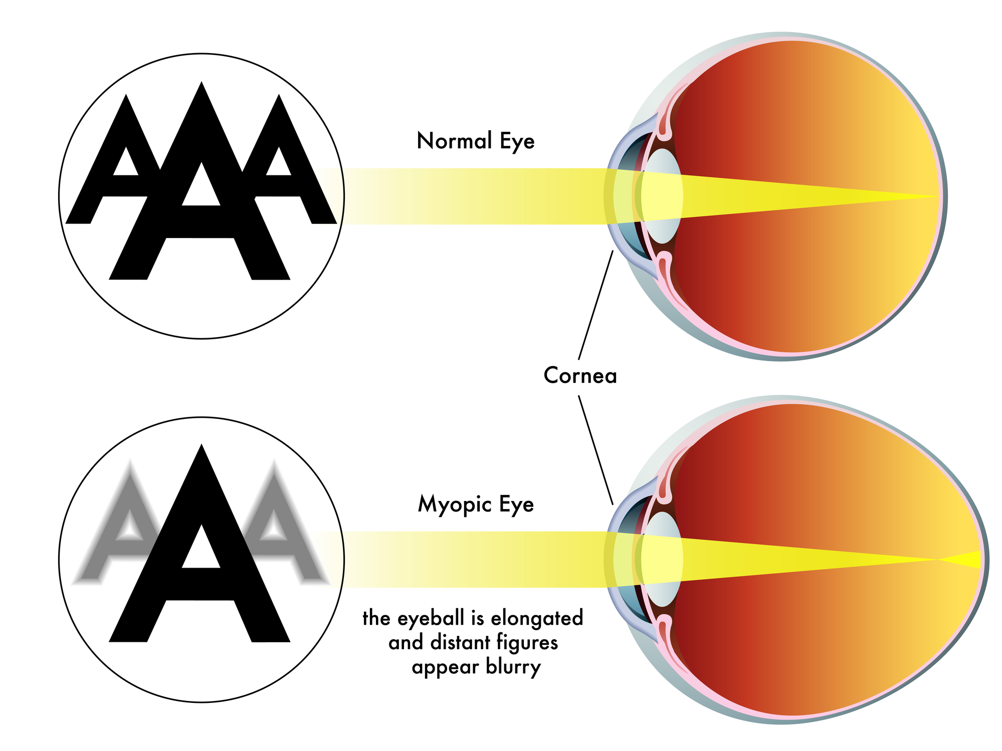
Prescription glasses online offer convenience, variety, and affordability—just ensure accurate measurements and trusted sellers for satisfaction.
 By
By
Lasers in medicine enhance surgical precision, reduce recovery times, and expand treatment options across multiple specialties.
 By
By
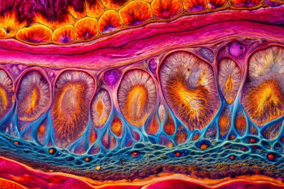
Advanced dermatological imaging provides detailed insights into skin layers, enhancing early disease detection, treatment accuracy, and overall patient care significantly.
 By
By
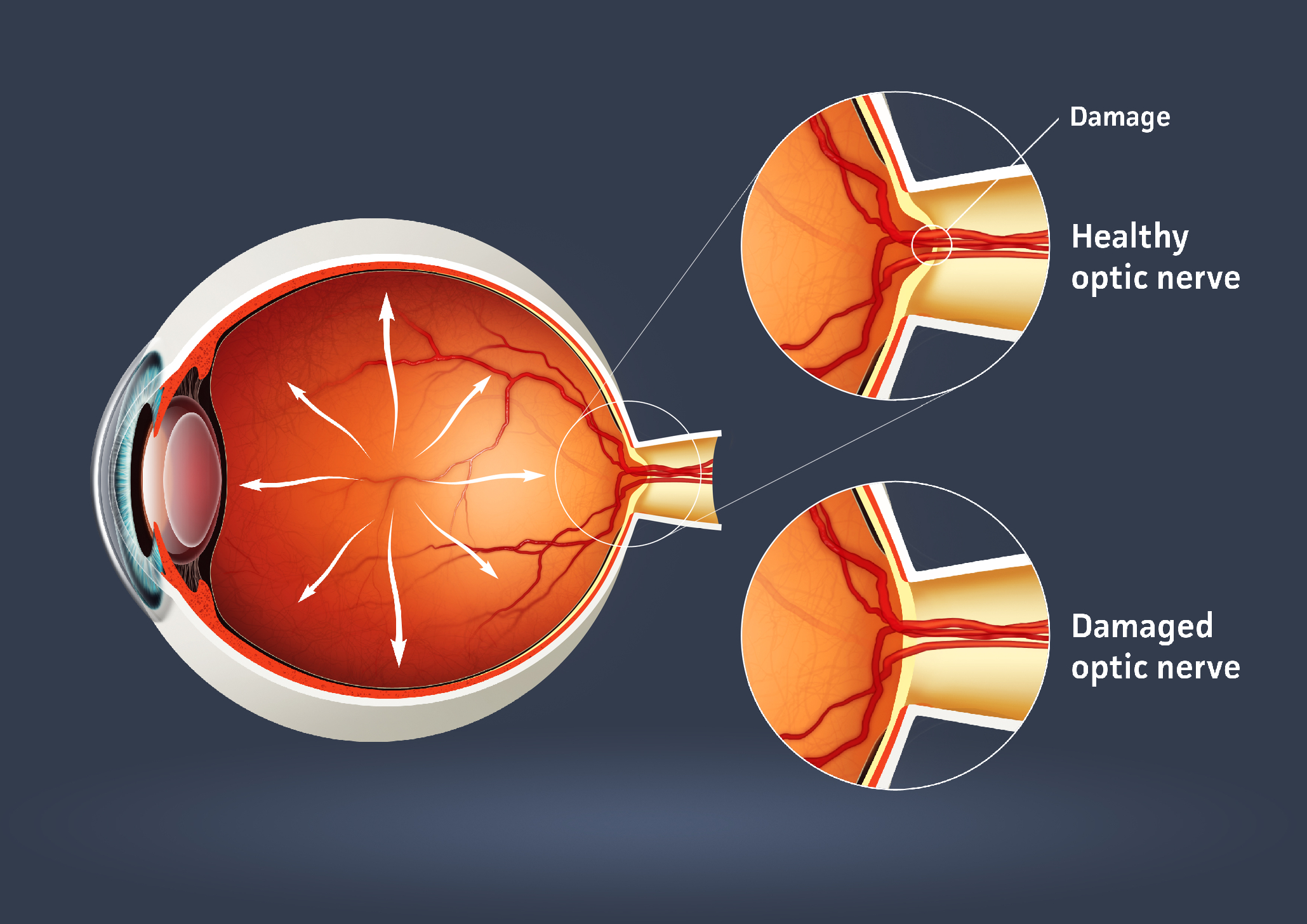
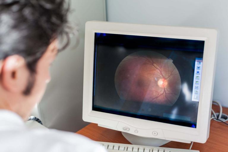
Medical imaging, crucial for optic nerve disorders, has evolved with technologies like MRI and OCT. Image for illustration only. Person depicted is a model.
 By
By
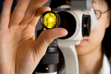
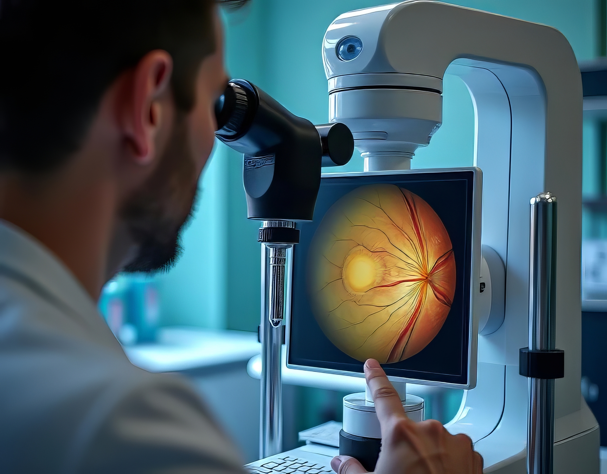
Since the 1800s, optic disc photography has been considered the gold standard for optic nerve evaluation. Image for illustration only. Person depicted is a model.