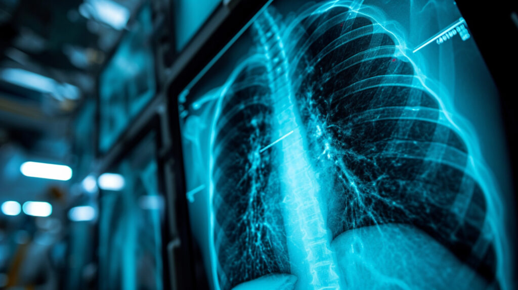
Deep learning predicts paediatric age from chest X-rays
 By
By
Explore the potential of paediatric age estimation through deep learning and chest X-rays for accurate age assessment.
X-ray attenuation modelling is a crucial aspect of medical imaging, materials science, and industrial radiography. It describes the reduction in X-ray intensity as the beam passes through a material, governed by fundamental interactions such as photoelectric absorption, Compton scattering, and, at higher energies, pair production. Understanding and accurately modelling X-ray attenuation is essential for optimising imaging techniques, improving radiation safety, and enhancing material characterisation.
Fundamentals of X-ray Attenuation
The attenuation of X-rays is mathematically described by the Beer-Lambert law:
I = I0e−μx
Where I is the transmitted intensity, I0 is the incident intensity, μ is the linear attenuation coefficient, and x is the thickness of the material. The attenuation coefficient, μ, depends on the material’s density and atomic composition, as well as the X-ray photon energy.
Two primary mechanisms contribute to X-ray attenuation in most diagnostic and industrial applications:
At very high photon energies (above 1.022 MeV), pair production can occur, where the photon transforms into an electron-positron pair, though this is rare in diagnostic imaging.
Modelling Techniques
Accurate modelling of X-ray attenuation requires detailed knowledge of material properties and photon interactions. Several approaches exist:
Applications and Challenges
X-ray attenuation modelling plays a key role in computed tomography (CT), where accurate attenuation coefficients enable tissue differentiation. It is also essential in non-destructive testing, security screening, and radiation shielding design. The main challenge lies in modelling heterogeneous structures, where attenuation varies spatially. Advances in AI-driven modelling and improved Monte Carlo techniques continue to enhance accuracy and efficiency in this field.
home »
By
Explore the potential of paediatric age estimation through deep learning and chest X-rays for accurate age assessment.
 By
By
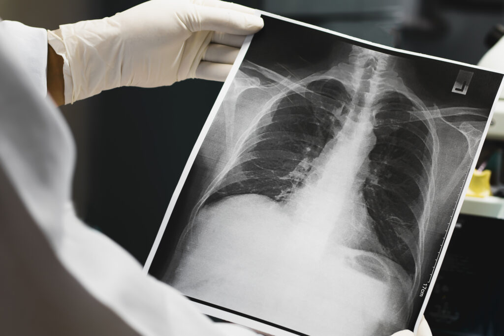
Discover the journey of X-rays in medicine through a real patient case and their importance in clinical diagnostics.
 By
By
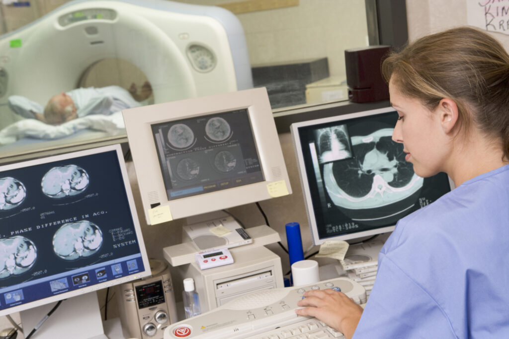
Understand the principles of CT imaging in clinical practice, including attenuation and dose management for accurate diagnoses. Image for illustration only. People depicted are models.
 By
By
Explore the evolution of imaging health and its impact on diagnosis, treatment, and prevention in modern medicine.
 By
By
Kilovoltage X-rays provide clear imaging contrasts, supporting precise diagnoses and effective treatment of superficial skin lesions.
 By
By
Diagnostic Imaging in Sports Medicine helps accurately diagnose injuries, guiding effective treatment and optimising athlete recovery timelines.