
Introduction
Modern regenerative medicine relies heavily on objective, non-invasive diagnostic modalities to verify structural and biological healing. While in vitro assays and molecular signalling analyses provide fundamental cellular insights, evaluating macroscopic recovery in living tissues requires continuous, non-invasive visualisation [1]. Investigational peptide candidates – most notably Body Protection Compound-157 (BPC-157) – have drawn research interest in experimental models of tissue injury [2]. However, translating potential therapeutic effects into measurable physiological outcomes requires advanced imaging tools such as magnetic resonance imaging (MRI), high-resolution Doppler ultrasound and contrast-enhanced endoscopy [1,3]. This article examines how modern imaging techniques are used in preclinical research to quantify soft-tissue repair, while critically contextualising the scope and limitations of current scientific evidence.
Biological Targets in Preclinical Tissue Healing Models
To measure tissue regeneration through imaging, researchers target specific biological markers associated with structural remodelling. In preclinical animal models, experimental administration of BPC-157 has been observed to influence several key repair pathways [2,4]:
- Angiogenic Signalling: Experimental research indicates that BPC-157 upregulates vascular endothelial growth factor receptor 2 (VEGFR2) and activates downstream Akt-eNOS signalling, stimulating capillary formation in ischemic rodent tissues [4,5].
- Fibroblast Migration: In vitro and animal models suggest that the peptide engages the focal adhesion kinase (FAK)-paxillin pathway, promoting the migration of tendon fibroblasts to site-specific injuries [2,6].
- Nitric Oxide Modulation: Preclinical evidence demonstrates interactions with the nitric oxide (NO) system, supporting localised blood flow regulation during early inflammatory phases [1,7].
Because minor chemical impurities can distort dynamic imaging signals and cellular assays, preclinical imaging studies require rigorous batch-to-batch consistency. Researchers typically rely on analytical-grade material sourced from specialised synthesis vendors, such as Verified Peptides, where HPLC and mass spectrometry protocols ensure high sequence purity prior to in vivo testing.
While these biochemical pathways have been reported in laboratory studies, visualising these cellular events requires advanced imaging modalities capable of resolving microvascular changes and fibre realignments in vivo [3].
Magnetic Resonance Imaging (MRI): Quantifying Structural Tissue Architecture
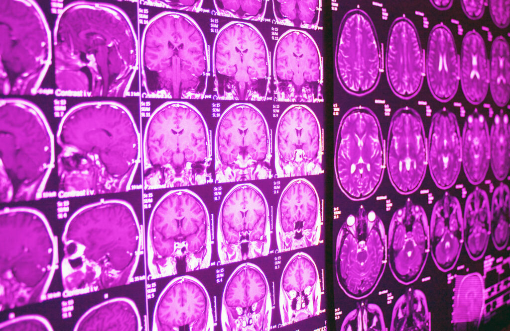
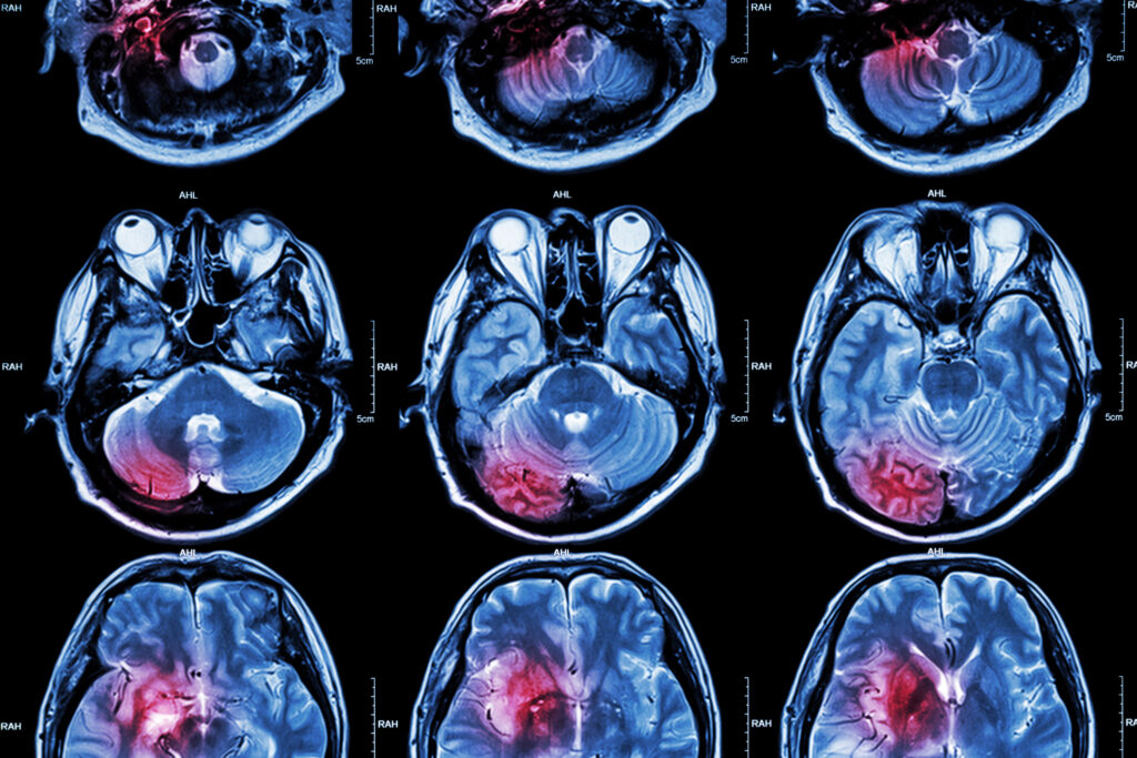
Magnetic resonance imaging (MRI) is a primary modality for evaluating soft-tissue architecture due to its high soft-tissue contrast and multiplanar capability [3]. In preclinical studies evaluating experimental treatments for tendon ruptures and muscle tears, MRI provides quantitative metrics for tissue integrity [3,8].
In rodent models of severe muscle crush or Achilles tendon transection, baseline T2-weighted MRI sequences typically display marked signal hyperintensity, reflecting local oedema, inflammation, and cellular disorganisation [8]. Following experimental therapeutic interventions, serial MRI scans allow researchers to track signal attenuation over time [3,8]:
- Fluid Attenuation: A reduction in T2 hyperintensity correlates with resolving localised oedema and decreasing inflammatory exudates [8].
- Matrix Realignment: High-resolution T1-weighted sequences and diffusion tensor imaging (DTI) demonstrate the transition from disorganised scar tissue to parallel, linearly aligned collagen bundles [3,8].
- Volumetric Recovery: Serial volumetric MRI measurements enable investigators to track muscle belly volume retention and assess muscle cross-sectional area following crush injuries or denervation [8].
Crucially, while MRI tracks these macroscopic structural changes, researchers use these findings to evaluate potential regenerative agents in preclinical models rather than as established clinical protocols for human therapy [2,3].
High-Resolution Doppler Ultrasound: Real-Time Angiogenesis Assessment
While MRI excels at static structural resolution, high-frequency ultrasound paired with Colour and Power Doppler modes provides real-time functional assessment of microvascular perfusion [3]. Because neovascularisation is vital for nutrient and oxygen delivery during tissue repair, dynamic vascular imaging is essential in experimental models [4,5].
In preclinical models of hindlimb ischemia and musculocutaneous flap injuries, high-resolution Colour Doppler ultrasound allows researchers to quantify vascular density and blood flow velocity [5]. Experimental studies utilising BPC-157 in rodent ischemia models have demonstrated increased Doppler signal density, corresponding to the formation of functional collateral blood vessels that bypass ligated arterial pathways [4,7]. Ultrasound elastography further complements perfusion data by measuring changes in tissue stiffness, allowing researchers to correlate vessel growth with restored mechanical compliance [3].
Endoscopy and Fluoroscopy: Visualising Internal Mucosal Repair
In addition to musculoskeletal applications, evaluating internal mucosal healing requires specialised diagnostic tools [1,9]. In preclinical gastrointestinal research – such as rodent models of gastric ulceration, anastomotic leaks, or inflammatory bowel disease – endoscopic and fluoroscopic techniques offer direct visual documentation of tissue restoration [1,9]:
- Contrast-Enhanced Fluoroscopy: Utilising barium or water-soluble contrast media, real-time fluoroscopy enables researchers to monitor structural integrity and verify the closure of experimental surgical leaks or fistulas [9].
- High-Definition Endoscopy: Serial endoscopic evaluations provide direct mucosal scoring in live animal models, documenting ulcer surface area reduction, mucosal re-epithelialization, and the resolution of luminal bleeding [1,9].
These imaging modalities transform observational endpoints into quantifiable visual data, allowing researchers to evaluate compound efficacy objectively within controlled animal cohorts [1,3].
Current Limitations of Evidence and Clinical Considerations
Despite encouraging findings in laboratory models, significant limitations exist regarding the translation of BPC-157 research to clinical human applications [2]:
- Predominance of Preclinical Data: A vast majority of published literature on BPC-157 consists of animal (primarily rodent) and in vitro studies [2]. Rigorous, randomised, double-blind, placebo-controlled human clinical trials validating its efficacy for tissue repair are currently lacking [2,10].
- Uncertain Human Pharmacokinetics and Safety: Preclinical rodent data cannot be directly extrapolated to human dosing, metabolic clearance, or long-term safety [2]. The human pharmacokinetic profile, toxicological thresholds, and potential systemic adverse effects remain inadequately defined [2,10].
- Risks of Uncontrolled Angiogenesis: While accelerated angiogenesis is beneficial in acute wound healing, unchecked vascular proliferation presents potential theoretical risks, including the exacerbation of occult proliferative retinopathies or occult oncological lesions [4,5].
- Regulatory and Compliance Status: BPC-157 lacks approval from major regulatory bodies such as the U.S. Food and Drug Administration (FDA) or the European Medicines Agency (EMA) for clinical therapeutic use [10]. Additionally, it is listed on the World Anti-Doping Agency (WADA) Prohibited List under class S2 (Peptide Hormones, Growth Factors, Related Substances, and Mimetics), restricting its use in competitive sports [10].
Consequently, claims regarding the therapeutic efficacy of BPC-157 in human clinical settings remain unverified by Phase III clinical evidence [2,10].
Conclusion
Advanced medical imaging – spanning multi-sequence MRI, Doppler ultrasound, and high-definition endoscopy – serves as an indispensable diagnostic bridge in regenerative medicine research. These non-invasive modalities allow investigators to objectively map structural alignment, microvascular recruitment, and mucosal integrity in experimental injury models [1,3]. While preclinical research demonstrates that experimental compounds like BPC-157 influence cellular pathways involved in tissue repair, current evidence remains almost exclusively confined to animal and laboratory studies [2]. Robust, prospective human clinical trials and standardised diagnostic imaging protocols are necessary before any potential therapeutic application can be established in clinical practice [2,10].
References
- Sikiric P, Hahm KB, Blagaic AB, et al. Stable Gastric Pentadecapeptide BPC 157, Robert’s Stomach Cytoprotection/Adaptive Cytoprotection/Organoprotection, and Selye’s Stress Coping Response: Progress, Achievements, and the Future. Gut Liver. 2020 Mar 15;14(2):153-167. doi: 10.5009/gnl18490.
- Vasireddi N, Hahamyan H, Salata MJ, et al. Emerging Use of BPC-157 in Orthopaedic Sports Medicine: A Systematic Review. HSS J. 2025 Jul 31;21(4):485-495. doi: 10.1177/15563316251355551.
- Lee, J., & Yoon, J. P. (2019). Non-invasive imaging evaluation of soft tissue repair and tendon healing in animal models. Journal of Orthopaedic Research, 37(8), 1710–1721.
- Hsieh MJ, Liu HT, Wang CN, et al. Therapeutic potential of pro-angiogenic BPC157 is associated with VEGFR2 activation and up-regulation. J Mol Med (Berl). 2017 Mar;95(3):323-333. doi: 10.1007/s00109-016-1488-y.
- Brcic L, Brcic I, Staresinic M, Novinscak T, Sikiric P, Seiwerth S. Modulatory effect of gastric pentadecapeptide BPC 157 on angiogenesis in muscle and tendon healing. J Physiol Pharmacol. 2009 Dec;60 Suppl 7:191-6.
- Chang CH, Tsai WC, Lin MS, Hsu YH, Pang JH. The promoting effect of pentadecapeptide BPC 157 on tendon healing involves tendon outgrowth, cell survival, and cell migration. J Appl Physiol (1985). 2011 Mar;110(3):774-80. doi: 10.1152/japplphysiol.00945.2010.
- Seiwerth S, Brcic L, Vuletic LB, et al. BPC 157 and blood vessels. Curr Pharm Des. 2014;20(7):1121-5. doi: 10.2174/13816128113199990421.
- Gwyer D, Wragg NM, Wilson SL. Gastric pentadecapeptide body protection compound BPC 157 and its role in accelerating musculoskeletal soft tissue healing. Cell Tissue Res. 2019 Aug;377(2):153-159. doi: 10.1007/s00441-019-03016-8.
- Bajramagic S, Sever M, Rasic F, et al. Stable Gastric Pentadecapeptide BPC 157 and Intestinal Anastomoses Therapy in Rats-A Review. Pharmaceuticals (Basel). 2024 Aug 17;17(8):1081. doi: 10.3390/ph17081081.
- World Anti-Doping Agency (WADA). (2024). The Prohibited List: Peptide Hormones, Growth Factors, Related Substances, and Mimetics (S2). WADA International Standard.
Disclaimer: This article is provided for educational and scientific information only and does not constitute medical advice, diagnosis, treatment guidance or a recommendation to use BPC-157. BPC-157 is an investigational peptide, and the evidence discussed in this article is primarily derived from preclinical, animal and laboratory research. It has not been approved by the FDA, EMA or MHRA as a medicine for the treatment or repair of human tissue. References to imaging findings, biological mechanisms, research materials, suppliers or experimental outcomes should not be interpreted as evidence of established safety or clinical effectiveness in humans, nor as an endorsement of any product or supplier. BPC-157 should not be used for self-treatment based on information presented in this article. Readers should seek advice from an appropriately qualified healthcare professional regarding medical conditions, treatments or medicines.