You hear the phrase ‘generative AI enables medical image segmentation in ultra-low data regimes’, and it sounds like something that’s meant for a research lab, not something that has an effect on real-world medicine.
But it does.
Actually, you’ve already seen the problem it tries to solve without you realising it.
Here’s a quick real-world example: a radiologist staring at a scan where only a handful of similar cases exist, and the software that’s meant to help can’t recognise what it’s looking at. When you have a tiny dataset, every pixel is important, and every mistake is a big deal.
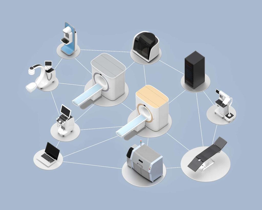
AI thrives on a scale, but you probably already know that. Medicine, however, doesn’t have the same luxury because there aren’t thousands of photographs of rare tumours. Hospitals don’t share their data just like that, and labelling one MRI slice can take longer than reading an entire chest CT.
For you, that means that you’re trying to make sense of important scans, but you don’t have enough examples to teach an AI model what ‘normal’ or ‘abnormal’ even looks like.
This is where generative models turn the entire situation upside down, so let’s get into more details on that.
Why Medical Image Segmentation Fails in Low-Data Scenarios
Approx. 25-30 million people in the U.S. are affected by 6,800+ rare diseases each year, with only a fraction having documented imaging cases.
It’s always the same problem with traditional segmentation models: they need a lot of labelled images to learn anything meaningful.
A network like U–Net becomes unreliable when you only feed it a small handful of examples because, instead of trying to understand the underlying anatomy, it just starts memorising scans. That’s the reason why a model trained in one hospital usually falls apart the moment you test it on images from a different scanner or a different group of patients.
And rare conditions make this problem even worse.
One single radiologist-produced segmentation can take a dozen minutes per slice; an MRI can have 100+ slices, which leads to multiple hours of expert-level work for each case.
Some tumours and abnormalities show up in only a few cases a year, and even when they do appear, they hardly ever look the same twice. So not only do you have very little data, but you also have a lot of variation.
This basically means that the model doesn’t see enough patterns of what it’s designed to look for.
To add to that, every labelled mask comes from a radiologist who outlines structures pixel by pixel – as you can imagine, that takes a lot of time and money. And even if you’ve got the resources, the labelled masks will vary from one expert to another, which might negatively affect consistency or require additional editing work.
The inconsistencies are also affected by other factors (e.g., scan quality, slice thickness, noise levels, differences in protocol, etc.), which ultimately results in a dataset that becomes very small after you filter everything you cannot use.
How Generative AI Overcomes Ultra-Low-Data Constraints
When you don’t have a lot of labelled scans to work with, you turn to generative models so you can expand that dataset without waiting for more patients, more annotations, etc. You don’t try to squeeze every last drop of information out of a tiny set of MRIs or CTs and, instead, you let the model fill in the gaps.
It creates new examples and variations that help the segmentation network understand what real anatomy looks like, even when the actual dataset is small.
Exactly 755 medical devices were included in the cohort of FDA-approved AI/ML-based devices.
Synthetic Image Generation
Generative models like diffusion networks and GANs do great at creating artificial medical images that look and behave like the real thing. They can generate CT, MRI, and ultrasound scans with realistic textures and anatomical structures, which gives you a lot more material to train on than the original dataset provides.
3DINO, a self-supervised learning (SSL) framework designed for 3D medical imaging (MRI, CT, PET), is able to reduce labelled data needs by 4- 10x with quantifiable improvement to overall performance, even on the ultra-low-data regime (approx. 10 scans).
These synthetic images don’t have to be random, either.
The models can simulate slight variations in anatomy, add scanner noise, adjust contrast, and recreate situations that the limited dataset never captured.
Another plus is that synthetic images often come with built-in segmentation masks when the model is trained to output both the image and the label at the same time.
Generative Data Augmentation
Generative augmentation doesn’t work the same as the usual flipping or zooming tricks.
When you stretch/distort existing images, you’re running into the issue of the image not being anatomically correct.
This issue can be resolved using generative AI models, which can be used to create multiple new versions of the original image. And if the situation requires it, small changes (e.g., tissue density, lesion size, layered segments, contrast, motion blur to mimic scans, the model can do variations, etc) can still be made to each of the generated variations of that original image.
The goal is to get a greater variety of examples that the segmentation sees, but without introducing anything anatomically inaccurate.
You see a similar problem in other fields that deal with limited data, like cases that involve parking lot slip and fall injuries, industrial equipment-related accidents, chemical exposure incidents, low-volume product defect injuries, etc.
Because of the nature of these types of injuries, where high variability is common, in each of these situations, you need more examples or supplemented data before any pattern can become clear.
Self-Supervised and Foundation-Model Pretraining
Foundation models make low-data training easier because they learn the basics of medical anatomy before you ever fine-tune them.
3DINO-ViT, a model produced by training a Vision Transformer (ViT-Large) using the 3DINO framework, achieved segmentation performance that’s comparable to models trained on 100% of the labelled data while it used less than 50% of the labelled dataset during its fine-tuning.
They train on huge sets of unlabeled scans and pick up general features like organ boundaries and common intensity levels. Methods like masked autoencoders and contrastive pretraining help the model learn these features without any manual labels.
When you add your small labelled dataset later, the model already understands the structure of medical images, so it doesn’t need as many annotated examples to perform well on your segmentation task.
Pseudo-Labelling and Semi-Supervised Learning
Thanks to pseudo-labelling, you’re able to use unlabeled scans without turning the whole process into a manual job for radiologists.
First, the model creates rough segmentation masks on its own. Then you have an expert review a small portion of those predictions and correct the ones that are the most important. These corrected masks guide the next round of training, and the model gradually improves because it learns from both verified and unverified predictions.
The result is a large, unlabeled dataset being turned into something useful with less expert labour needed.
Over time, you end up with a stronger, more reliable segmentation model even if all you had to start with was a small number of labelled images.
Conclusion
This entire problem really comes down to one thing, and that’s the fact that medical imaging rarely gives you the luxury of working with endless examples.
You work with what you have and, more often than not, it’s not much. This meant that your segmentation model hit a wall fast, but generative AI changes that in a way that seems almost unfair. In a good way, though.
And this change opens a door for segmentation tools that are faster to build and that adapt more easily.
But most importantly, they’re far more reliable than anything you could train the old way.
Disclaimer
The information provided in this article is intended for educational and informational purposes only. It should not be interpreted as clinical advice, diagnostic guidance, or a substitute for professional medical judgement. Any tools, techniques, or AI-driven methods described here may not be suitable for every clinical setting and should only be applied by qualified professionals who can evaluate their appropriateness within the context of specific medical, regulatory, and ethical requirements.
Generative AI systems used for medical imaging tasks are subject to ongoing research and development. Performance can vary depending on data quality, hardware, software configuration, and clinical workflow. Readers should not assume that examples, models, or outcomes discussed in this article will produce identical results in their own environment.
Open MedScience does not endorse or guarantee any particular technology, product, or model referenced, and bears no responsibility for decisions made based on the content of this publication. Clinicians, researchers, and organisations should rely on their own expertise, independent testing, and regulatory guidance before deploying or integrating any AI-based medical imaging solutions.
home » blog » medical technologies »