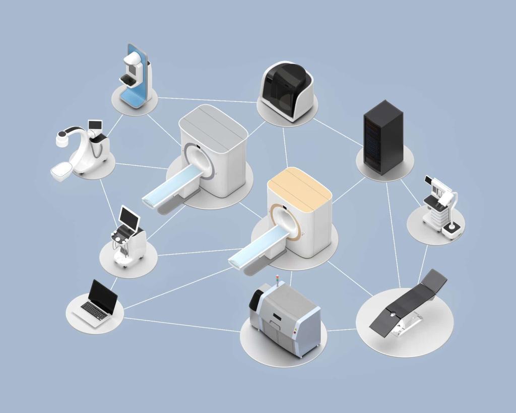
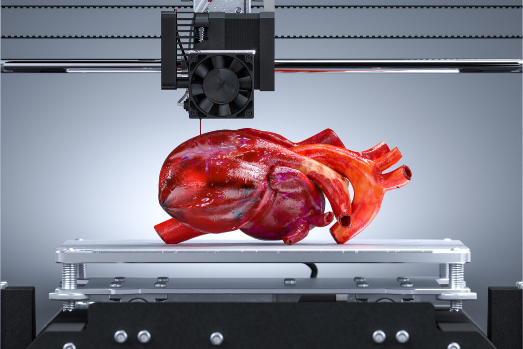
Summary: Advanced visualisation techniques, including 3D rendering, virtual reality (VR), and augmented reality (AR), are revolutionising the practice of radiology by enhancing how clinicians interpret and interact with medical images. These technologies provide highly detailed and immersive perspectives, enabling specialists to improve diagnostic precision, refine surgical planning, and enhance patient education. This article explores the historical evolution of advanced visualisation, examines 3D imaging, VR, and AR in detail, and discusses their impacts on patient care, education, and the overall progression of radiology.
Keywords: Radiology; 3D visualisation; Virtual reality; Augmented reality; Medical imaging; Patient education.
Introduction to Radiology
Over the past several decades, radiology has been at the forefront of medical innovation. Imaging technologies, from the earliest days of X-rays and computed tomography (CT) to more recent advancements like magnetic resonance imaging (MRI), have provided healthcare professionals with new ways to investigate and interpret the body’s structures. In the modern era, the leap forward in computing power has allowed radiologists not only to capture images but also to manipulate and view them in novel forms. Traditional two-dimensional (2D) images on film or screens are no longer the final word in imaging. Instead, a new dimension of possibility has arrived in the shape of advanced visualisation methods, including 3D rendering, VR, and AR.
Although the role of advanced visualisation in radiology might once have been considered aspirational, it is now rapidly transitioning into routine use. Hospitals and diagnostic centres around the world are adopting these advanced techniques to achieve better patient outcomes and more accurate diagnoses. The heightened clarity and immersion offered by 3D, VR, and AR technologies enhance the radiologist’s understanding of organs, tissues, and pathologies, facilitating more informed treatment recommendations. These tools also aid surgeons by providing thorough preoperative assessments and simulations of complex anatomical structures.
Moreover, advanced visualisation extends its benefits beyond purely diagnostic applications. Patient education gains considerable advantages when patients can see their anatomy in vivid digital form, allowing them to grasp their conditions and the rationale behind proposed treatments. Medical students also benefit from these tools, as they can rotate, enlarge, and virtually explore anatomical structures without the need for traditional cadaveric dissection or 2D diagrams.
By understanding how 3D rendering, VR, and AR are used in clinical scenarios, healthcare providers can refine their approaches and ensure safer and more effective patient care. Ultimately, the synthesis of these advanced methods forms a new paradigm in radiology, altering how medical professionals see, analyse, and plan interventions in human anatomy.
Historical Evolution of Visualisation in Radiology
Radiology’s long history can be traced back to the discovery of X-rays by Wilhelm Conrad Röntgen in 1895. By enabling physicians to see beneath the skin, Röntgen’s discovery opened unprecedented diagnostic and research possibilities. Early radiology was limited to plain film X-rays, which, although helpful, provided only flat, 2D shadows of the body’s internal features. These images were inherently simplistic and often required extensive interpretation skills to translate the shadows into a coherent understanding of three-dimensional anatomical relationships.
As film radiography became a standard practice, researchers sought ways to extract more detailed information. The introduction of contrast agents, such as barium, highlighted specific structures, making them more distinct on X-ray film. This incremental improvement was followed by the invention of techniques like fluoroscopy, which provided real-time 2D images yet remained limited in-depth perception and clarity for complex anatomical regions.
The Shift to Digital Imaging
The real revolution in radiology began in the 1970s and 1980s with the development of CT scans and MRI. Both these modalities brought new layers of detail to the imaging process. CT scans utilise sophisticated algorithms to assemble cross-sectional images from multiple X-ray measurements, while MRI employs magnetic fields and radio waves to derive slices of data with exceptional soft-tissue contrast. As computer hardware advanced, it became easier to process and store these digital slices, laying the groundwork for three-dimensional reconfigurations of anatomical structures.
With digital imaging, radiology moved further into computer-based interpretation. Technological innovators recognised that stacking hundreds of CT or MRI slices could yield 3D representations. In the 1990s, some institutions began experimenting with so-called “volume rendering.” This technique used algorithms to construct a volumetric model of the scanned area, potentially rotating it and slicing it in ways that 2D images could not match.
Emergence of Advanced Visualisation
Once powerful computing machines became more affordable, advanced visualisation programs were refined to present complex 3D views in real-time. Radiology workstations enabled radiologists to zoom in, rotate, and virtually cut through 3D volumes, transforming how specialists read and interpret scans. These early forms of 3D visualisation primarily relied on external monitors. However, the groundwork was being laid for immersive and interactive technologies like VR and AR.
The advent of modern VR headsets and AR-enabled devices in the consumer market, particularly around the early 2010s, created an opportunity for radiology. Researchers and clinical innovators realised these devices could be adapted for medical imaging. The combination of established 3D rendering techniques with VR and AR hardware signalled a new chapter in radiological practice. This integration not only offered improved situational awareness but also facilitated more intuitive interactions with medical data.
3D Imaging in Radiology
The simplest form of advanced visualisation in radiology is 3D rendering. Modern scanners can capture enormous amounts of data in a short time. For example, a high-resolution CT scan can produce hundreds or even thousands of cross-sectional slices of a single region of the body. Each slice provides a thin “layer” of information. When stitched together, these layers can be used to construct a digitally rendered 3D model.
Common methods of 3D rendering include surface rendering and volume rendering. Surface rendering uses an outline of the scanned object, emphasising the boundaries between structures, such as bones or blood vessels. Volume rendering is a more thorough process that accounts for densities and transparencies within tissues, creating a richer visual experience. This is especially helpful when viewing soft tissues and trying to identify abnormalities concealed by overlapping structures in 2D.
Applications of 3D Imaging
In practical radiology, 3D imaging has a host of applications. For instance, 3D reconstructions of blood vessels in the brain can help detect and illustrate aneurysms, guiding endovascular interventions. Orthopaedic surgeons can inspect intricate bone fractures or plan surgeries more easily when they have an accurate 3D representation of a joint or a complex fracture site. Cardiothoracic specialists can investigate structural heart issues and visualise blood flow through 3D models reconstructed from CT or MRI data.
A key advantage of 3D imaging is the ability to re-slice data in any plane. Traditional axial views (top-to-bottom) are sometimes insufficient for detecting subtle anomalies that become more obvious in the coronal (front-to-back) or sagittal (side-to-side) planes. With 3D imaging, radiologists can explore these planes interactively, adjusting windows and levels to accentuate certain tissue types or densities. This flexibility reduces the risk of missing small lesions that might be invisible or inconspicuous in a single plane.
Benefits for Diagnosis and Treatment
For diagnosis, 3D imaging offers a clearer depiction of relationships between tumours and adjacent structures, such as blood vessels and nerves. This fosters more accurate staging of diseases, such as cancer, and informs clinical decision-making about whether surgery, chemotherapy, radiotherapy, or a combined approach is the most suitable. In addition, 3D visualisation assists in planning the surgical approach to remove a tumour while safeguarding essential blood vessels and nerves.
Beyond tumour assessment, 3D models help in evaluating congenital anomalies. Surgeons operating on children with congenital heart defects, for example, gain a far better grasp of the anatomical layout through 3D reconstructions than with flat images alone. This improved understanding shortens operating times, diminishes the risk of complications, and leads to more effective procedures.
Virtual Reality in Radiology
VR entails creating a simulated environment that users can experience through specialised headsets. These headsets typically contain two small screens—one for each eye—and motion sensors that track head movements. When a user moves their head, the viewpoint within the virtual environment shifts accordingly, offering a sense of immersion not possible with standard monitors.
In medical imaging, VR platforms use the same raw data (from CT, MRI, or other scans) but present it in an interactive 3D space. Rather than merely looking at a 3D model on a flat screen, the user can enter a representation of the body, walking or flying through a patient’s anatomy in a way reminiscent of high-end gaming experiences but with scientific data as the foundation.
Advantages of VR for Radiologists
- Immersion and Depth Perception: VR delivers a direct sense of scale and spatial orientation. A lesion’s exact size in relation to surrounding structures can be viewed in a more natural way, which is helpful for complex surgical planning.
- Improved Collaboration: VR systems can facilitate multi-user sessions. Surgeons, radiologists, and other healthcare professionals from different departments or even from different geographic locations can enter the same virtual environment and examine the data concurrently. This fosters better communication, reduces misunderstandings, and speeds up the decision-making process.
- Interactivity and Real-time Updates: Advanced VR platforms permit real-time editing of the visualised data. For instance, radiologists can highlight specific areas, add or remove layers, or annotate regions of concern while discussing with colleagues. These annotated VR sessions can then be saved for later use.
Clinical and Research Applications of VR
Early adopters of VR in radiology have highlighted its usefulness in neurosurgery. Brain tumours located near sensitive structures demand a thorough plan to minimise damage to healthy tissue. With VR, neurosurgeons can simulate different approaches to a tumour, examining potential risks associated with each route. This method not only refines surgical techniques but can also reduce operating times and complications.
VR-based radiology is equally valuable in fields like cardiac surgery, orthopaedics, and maxillofacial surgery. A maxillofacial surgeon, for instance, can navigate a patient’s jaw, zooming in on the angle of a fracture and determining the best way to place plates or screws. Similarly, a cardiac surgeon might explore the geometry of heart valves or major vessels within a virtual environment, making decisions about valve repairs or replacements.
Additionally, researchers use VR simulations of medical images to develop new approaches for diagnosing and treating diseases. Some studies investigate whether the immersive qualities of VR allow for earlier detection of subtle lesions. Others explore how VR data might be integrated with robotic surgeries, enabling surgeons to see the patient’s anatomy from multiple angles while operating through robotic arms.
Augmented Reality in Radiology
While VR immerses a user in a completely computer-generated environment, AR overlays digital information onto the real world. AR systems often use transparent headsets or handheld devices (like tablets or smartphones) to project 3D information onto the user’s view of the physical space. The user still sees the real environment but with digital elements superimposed.
In the radiological context, AR merges real-time patient images with pre-acquired scans. For example, an interventional radiologist wearing AR glasses might look at a patient’s body and see an overlay of the internal organs’ 3D representation, guiding needle placement or catheter navigation.
AR Devices and Software
Several AR devices have entered the market, some focusing on consumer applications and others specifically designed for surgical or medical uses. These specialised devices incorporate depth sensors, cameras, and software algorithms that track the user’s position and orientation. By mapping the user’s environment in real-time, the AR headset can accurately align 3D medical data with the physical patient.
Crucial to AR in radiology is the registration process. The 3D model derived from scans must precisely match the patient’s actual anatomy, requiring careful calibration. Minor errors in alignment can lead to an incorrect understanding of the patient’s internal structures and compromise patient safety. Therefore, robust software solutions that handle real-time updates of posture, movement, and perspective are vital for successful AR implementation.
Clinical Applications and Benefits
- Image-guided Interventions: AR can improve image-guided procedures, such as biopsies, tumour ablations, and vascular interventions. By overlaying the patient’s anatomy onto the clinician’s field of view, AR helps the radiologist or surgeon pinpoint targets with greater precision, potentially reducing the duration of procedures and minimising complications.
- Surgical Navigation: Surgeons often rely on reference points—landmarks in the anatomy—along with 2D slices from CT or MRI scans for guidance. AR takes this one step further by displaying crucial information right on the surgical field. This is especially valuable in orthopaedic procedures, where a small misalignment in a joint replacement surgery can affect long-term patient outcomes.
- Patient Education: When explaining procedures, such as how an implant will be placed or how a tumour is positioned, an AR visualisation can offer immediate clarity. Patients who can see a digital overlay of their body’s internal structures during a consultation are more likely to grasp the condition and commit to the proposed plan.
Limitations and Future Opportunities
AR, though promising, faces limitations. The hardware, including headsets, can be bulky and might cause discomfort during longer procedures. Calibration and registration can be time-consuming, and the technology may struggle to adapt in real-time if the patient shifts position. Nonetheless, ongoing advances in device miniaturisation, software algorithms, and sensor accuracy will address many of these limitations. As computing power continues to evolve, we can anticipate AR solutions becoming more user-friendly, accurate, and integrated into routine clinical workflows.
Transforming Patient Education and Engagement
One of the most profound changes brought by advanced visualisation technologies is in patient education and engagement. Traditional methods of discussing medical conditions often involve static images or abstract explanations. For the average patient without medical training, it can be challenging to visualise an organ’s shape or a tumour’s location using 2D drawings or textual descriptions.
Through 3D rendering, VR, and AR, patients gain a far better view of their internal anatomies. A detailed 3D model presented on a workstation or a VR headset can demonstrate the patient’s actual condition rather than a generic example. This personalisation fosters greater trust and reduces anxiety because patients feel they are seeing the real scenario.
Virtual Consultations and Interactive Demos
Some institutions are experimenting with virtual consultations where patients, wearing VR headsets, can “tour” the inside of their own bodies alongside a medical professional. In these sessions, the clinician explains how specific structures appear and pinpoints any identified issues. Interactivity—allowing the patient to zoom in, rotate the model, or highlight areas of interest—empowers patients to ask informed questions.
This approach is particularly meaningful for patients facing major or complex surgeries. For instance, before undergoing a cardiac bypass, seeing a 3D model of the heart and the affected vessels can make the entire process more understandable. Patients often report feeling less apprehensive and more involved in decisions when they have visually absorbed how the procedure will address their particular issue.
AR Aids in Demonstrating Procedures
AR can take patient education a step further by superimposing digital elements on a patient’s own body. During consultations, a clinician might use an AR-compatible device to project a digital model of an implant or a surgical path onto the patient’s torso, making it crystal clear where incisions will be made and how the implant will fit in place. This immediate visual representation reinforces transparency and trust.
Additionally, patients can interact with AR or VR modules outside clinical consultations. Hospitals and clinics can develop or adopt existing educational software that simulates common procedures or pathologies. This self-guided learning, done at home or in waiting areas, equips patients with knowledge that can ease the stress often accompanying a serious health condition.
Impact on Medical Education and Training
Medical education has always depended heavily on teaching anatomy through cadaver dissection and 2D atlases. While these remain valuable learning tools, advanced visualisation offers an alternative or complementary route. VR anatomy labs are increasingly being introduced into medical schools, enabling students to interact with hyper-realistic models of organs, vessels, and tissues. They can explore the body layer by layer, remove muscles to expose deeper structures or simulate pathologies to understand how diseases alter normal anatomy.
In AR-based anatomy teaching, students can view each other wearing headsets that overlay floating anatomical structures in the classroom. This group learning environment encourages collaboration and helps reinforce key concepts, as students can point, label, and discuss different structures in real-time.
Surgical Simulation and Skill Development
Surgeons-in-training benefit immensely from advanced visualisation. Traditional surgical training requires them to learn in operating theatres under the supervision of experienced surgeons. Although this remains indispensable, VR-based simulators allow trainees to practise procedures repeatedly, refining techniques without risking patient safety. These platforms provide realistic haptic feedback and measure performance through metrics such as precision, speed, and the amount of tissue “damage” inflicted during virtual operations.
Complex procedures—neurosurgery, for instance—can be planned and practised in a virtual environment. Surgical residents who rehearse an operation several times in VR can enter the actual operating theatre feeling more confident and prepared. This can lead to shorter operating times and fewer errors, directly improving patient outcomes.
Remote Collaboration and Global Learning
An essential component of medical education and professional development is peer collaboration. Advanced visualisation extends this collaboration beyond the physical confines of hospitals or universities. Experts from around the world can gather in a virtual environment, examining the same 3D model of a patient’s anatomy. This promotes knowledge-sharing on a global scale, allowing medical trainees in resource-limited areas to learn from top experts.
Additionally, AR and VR can enable remote proctoring of surgical procedures. An experienced surgeon can watch a trainee operate from another city or country, offering real-time advice by manipulating and highlighting digital images superimposed over the trainee’s field of view. This synergy of education and technology holds promise for raising the standard of care in geographically isolated areas.
Pre-Surgical Planning and Interventions
Surgical interventions often hinge on the quality and detail of preoperative imaging. Traditional 2D images, while immensely valuable, do not always capture the complete three-dimensional character of pathology or its relationship to surrounding structures. Misunderstandings in pre-surgical planning can lead to suboptimal incisions, increased operative times, and a higher risk of complications.
Role of 3D, VR, and AR in Planning
3D reconstructions serve as a step up from standard scans. By creating detailed representations of organs, tumours, or vascular networks, they allow surgeons to map out surgical paths with enhanced precision. However, VR takes this concept further by immersing the surgeon in the patient’s anatomy. Interacting with the data in an immersive environment grants a more intuitive sense of depth and spatial arrangement.
For instance, in liver surgery, planning the resection margins near vital vessels is critical. A 3D or VR model can clarify exactly how much of the liver can be removed while still leaving enough healthy tissue. This knowledge informs the surgeon about the safest and most effective plan of attack.
AR can then be applied during the operation itself. By wearing an AR headset or using a display device, surgeons see the real operative field combined with the pre-planned 3D model. This fusion guides incisions and instrument placement with higher precision than is possible using only mental mapping from 2D images.
Interventions and Intraoperative Imaging
Advanced visualisation also impacts interventional radiology, which includes minimally invasive procedures guided by imaging. Procedures like ablating tumours with thermal probes or draining abscesses rely on accurate needle placement. AR-based needle navigation can shorten procedure times by displaying a projected path on the patient’s body. VR or 3D techniques may be used for planning how to route catheters through vascular structures without damaging fragile vessels.
Intraoperative imaging, such as real-time ultrasound fused with pre-acquired CT or MRI data, can be fed into a VR or AR system to update the display as the surgeon operates. This dynamic adjustment means that if a patient’s anatomy shifts or if a tumour is displaced during an operation, the surgeon can see these changes in real-time and adapt.
Technical Considerations and Implementation Challenges
Advanced visualisation in radiology depends heavily on hardware capable of handling large data sets. CT or MRI scans produce high-resolution images, and transforming these images into interactive 3D, VR, or AR formats demands substantial computing power. This includes GPUs for real-time rendering, high-performance CPUs, and large memory capacities to store volumetric data. VR or AR headsets add another layer of complexity, often needing motion-tracking cameras and precise sensors.
Healthcare facilities aiming to implement advanced visualisation must consider data storage infrastructure. Medical images can quickly consume terabytes of space, and the hospital’s network must support the rapid transfer of these files. Redundancy and backup systems are likewise vital to guard against data loss and preserve patient records.
Software Integration and Standards
On the software side, the diversity of file formats and imaging protocols (such as DICOM for medical scans) can create challenges. Systems must adhere to interoperability standards so that scans from different imaging devices can be integrated into a single 3D or VR environment. Developers must also ensure security and privacy, given the sensitive nature of medical data.
Professional organisations and consortia work on standardising protocols for data exchange and advanced visualisation tools. Hospitals, meanwhile, must evaluate the user-friendliness of any software platform they consider. If the system is too complex, radiologists and surgeons might be reluctant to incorporate it into their workflows. Comprehensive staff training and ongoing technical support are integral to the success of advanced visualisation implementation.
Cost and Return on Investment
Advanced visualisation systems can be expensive initially, requiring not only hardware and software but also training programs for staff. Institutions must evaluate the return on investment by looking at key metrics: improved diagnostic accuracy, shortened procedure times, decreased complication rates and enhanced patient satisfaction. Over time, these benefits can offset the costs by reducing readmissions, avoiding surgical errors, and streamlining clinical workflows.
Nonetheless, smaller clinics or underfunded hospitals might struggle to afford cutting-edge VR or AR setups. Efforts to make these technologies more accessible are ongoing, including the development of lower-cost headsets and open-source software solutions. Partnerships between technology companies and healthcare providers can also help reduce barriers to entry.
User Adoption and Training
Adopting new technology often requires a shift in mindset. Senior radiologists and surgeons who have practised with 2D imaging for decades might be hesitant to alter a tried-and-tested approach. Overcoming this resistance involves demonstrating clear benefits: fewer complications, more confident diagnoses, or simpler interprofessional communication.
Initial user training is critical. Hands-on workshops and integrated modules in medical school curricula can familiarise new generations of clinicians with advanced visualisation. As younger doctors become more comfortable with these tools, they can drive their adoption in day-to-day practice. Continuous education programmes for existing staff ensure that everyone remains updated about evolving techniques, new features, and best practices in advanced visualisation.
Ethical and Legal Implications
Digitally rendered anatomical models, especially those used in VR and AR, sometimes require remote data processing or cloud-based storage. As with any medical information, strong privacy regulations apply. Healthcare providers must ensure they comply with frameworks like the UK’s Data Protection Act and uphold standards set by regulatory bodies such as the General Medical Council.
Encryption and secure server environments help mitigate risks, but the potential for data breaches is always present. A leak or unauthorised access to VR-based surgical plans could reveal highly sensitive patient details. The ethical responsibility to protect patient confidentiality extends to these cutting-edge visualisation tools.
Responsibility and Liability
Advanced visualisation can enhance decision-making, but it also raises questions about responsibility and liability. If a surgeon relies heavily on a VR model to plan, a procedure and an unforeseen error occurs, how much of the fault lies with the technology provider versus the clinician? This question underscores the importance of thorough validation and regulatory approval for software that influences patient outcomes.
Likewise, AR systems must be meticulously tested to ensure that projected overlays align accurately with real anatomical structures. Small calibration errors can lead to significant complications during surgery. Healthcare providers must follow established guidelines and best practices when using advanced visualisation, keeping in mind that these tools are meant to supplement—not replace—the clinician’s expertise.
Informed Consent
When employing advanced visualisation for patient education or surgical planning, proper informed consent procedures must be followed. Patients should understand how their data will be used, stored, and visualised. This includes clarifying whether external collaborators or trainees can access their images in shared VR sessions. Ethical guidelines require that patients be informed of any technology’s experimental status and its potential limitations, ensuring they have a full picture before agreeing to its use.
Future Directions
One of the most exciting potential developments in advanced visualisation lies in the integration of artificial intelligence (AI). AI algorithms can assist in identifying lesions automatically, delineating tumour boundaries, or generating predictive analytics about disease progression. Incorporating these capabilities into VR or AR tools could result in real-time, AI-guided insights. As the clinician explores a 3D or VR model, the system could highlight suspicious tissue areas or suggest likely diagnoses based on statistical patterns.
Such AI-augmented visualisation might also assist in personalising treatment plans. By analysing a large dataset of past patient outcomes, the system could suggest the surgical route linked with the best success rates for a particular anatomy or pathology. Over time, as AI refines its predictions, radiologists and surgeons could see a new level of decision support.
Holographic Technologies
Holography, or the projection of 3D images into real space without needing specialised headsets, remains a nascent area but shows promise for medical imaging. If holographic projections become more precise and easier to generate, clinicians could collaborate around a table, viewing a floating 3D model of a patient’s anatomy without any gear. This would make group discussions more natural, reducing the barrier of personal headsets and strengthening interactive learning.
Remote Surgery and Telementoring
Though remote surgery has existed in limited forms, advanced visualisation and AR could push it to the next level. If a local team is physically present with the patient while a remote expert, equipped with AR displays, provides real-time guidance, procedures could be performed with near-equal expertise worldwide. High-speed internet connections, 5G and beyond, will likely make these scenarios more practical, turning telementoring into a routine component of surgical training and international medical collaboration.
Widening Adoption and Affordability
As with all emerging medical technologies, widespread adoption of advanced visualisation demands that costs drop and ease-of-use rises. Over the coming years, market competition and technological progress should lead to more affordable, user-friendly VR and AR systems. Educational institutions will increasingly build advanced visualisation methods into their curricula, guaranteeing that future generations of healthcare professionals will be adept at using these tools. This steady shift will help cement advanced visualisation as a standard component of radiological and surgical practice.
Conclusion
Advanced visualisation techniques are redefining the core of radiology. By allowing clinicians to view medical images in 3D or immerse themselves in VR and AR environments, these technologies transcend the limitations of 2D imaging. This shift has broad ramifications across diagnosis, patient education, surgical planning, and medical training. Surgeons can navigate anatomical structures with clarity that was once unattainable, while patients gain reassurance from visually transparent explanations of their conditions and potential interventions.
The path to full integration is not without barriers. Concerns around cost, hardware requirements, user adoption, and data security must be diligently addressed. Nevertheless, as technology rapidly evolves, these challenges are gradually being overcome. High-resolution imaging devices, robust software platforms, and collaborative frameworks have brought advanced visualisation from experimental novelty to emerging standard of care.
The future of radiology will likely see deeper synergy with fields such as AI and robotics. Predictive modelling, holographic displays, and remote-guided surgery are just a few of the frontiers that may be transformed by advanced visualisation. Students entering medical school today can look forward to a professional life enriched by these innovative tools, and established practitioners can harness them to enhance precision and efficiency.
Ultimately, the incorporation of 3D, VR, and AR into radiology represents a leap towards more nuanced patient care. It offers radiologists, surgeons, and patients alike a sharper perspective on the intricacies of the human body, fostering better outcomes and improved experiences. By embracing advanced visualisation, healthcare providers move closer to a future in which every patient benefits from a truly informed, cutting-edge approach to diagnosis and treatment.
Disclaimer
The content provided in this article is for informational and educational purposes only and is not intended as a substitute for professional medical advice, diagnosis, or treatment. While every effort has been made to ensure accuracy, Open Medscience makes no guarantees regarding the completeness, timeliness, or reliability of the information presented.
Technologies such as 3D visualisation, virtual reality (VR), and augmented reality (AR) continue to evolve rapidly. The clinical applications, regulatory approvals, and effectiveness of these technologies may vary by region, institution, and individual patient circumstances. Any references to clinical outcomes, procedures, or specific medical devices should not be construed as endorsements or recommendations.
Healthcare professionals should exercise their own clinical judgement and consult appropriate guidelines, regulatory bodies, and technology providers when considering the implementation of advanced visualisation methods in practice. Patients should not use this article as a basis for making healthcare decisions and are advised to consult qualified healthcare providers regarding any medical condition or treatment.
Open Medscience and the authors accept no liability for any loss or damage resulting from the use of information contained in this publication.
home » blog » medical technologies »


