3D Medical Imaging transforms diagnostics, treatment, and patient education through enhanced visualisation, precise localisation, and emerging AI integration.
The Emergence of 3D Medical Imaging: A Glimpse into the Future of Healthcare
3D Medical Imaging has radically transformed over the past century, driven by constant innovation and technological advancements. The advent of 3D imaging has marked a significant turning point in the field of radiology and nuclear medicine by providing healthcare professionals with a powerful and versatile tool for diagnosing, monitoring, and treating various disease states. The evolution from the rudimentary X-ray technology of the past to the sophisticated 3D imaging modalities employed today has truly transformed medicine, ultimately leading to improved patient outcomes.
This blog article will dive into the fascinating world of 3D medical imaging, examining the diverse range of technologies, their myriad applications, and numerous benefits leading to a promising future.
As 3D medical imaging continues to advance, imaging centres and diagnostic clinics generate increasing amounts of disposable items, including contrast packaging, single-use imaging components, and contaminated materials. Reliable medical waste disposal in Murfreesboro ensures these materials are handled and removed safely, allowing healthcare providers to focus on accurate diagnostics while maintaining clean, compliant clinical environments.
To fully comprehend the significance of 3D medical imaging, it is essential to understand its roots in earlier imaging technologies. Traditional X-ray technology, first discovered in 1895, provided a 2D representation of the human body’s internal structures. Although its limitations, X-ray technology was revolutionary for its time and laid the groundwork for more advanced imaging modalities, such as Computed Tomography (CT), Magnetic Resonance Imaging (MRI), and Ultrasound.
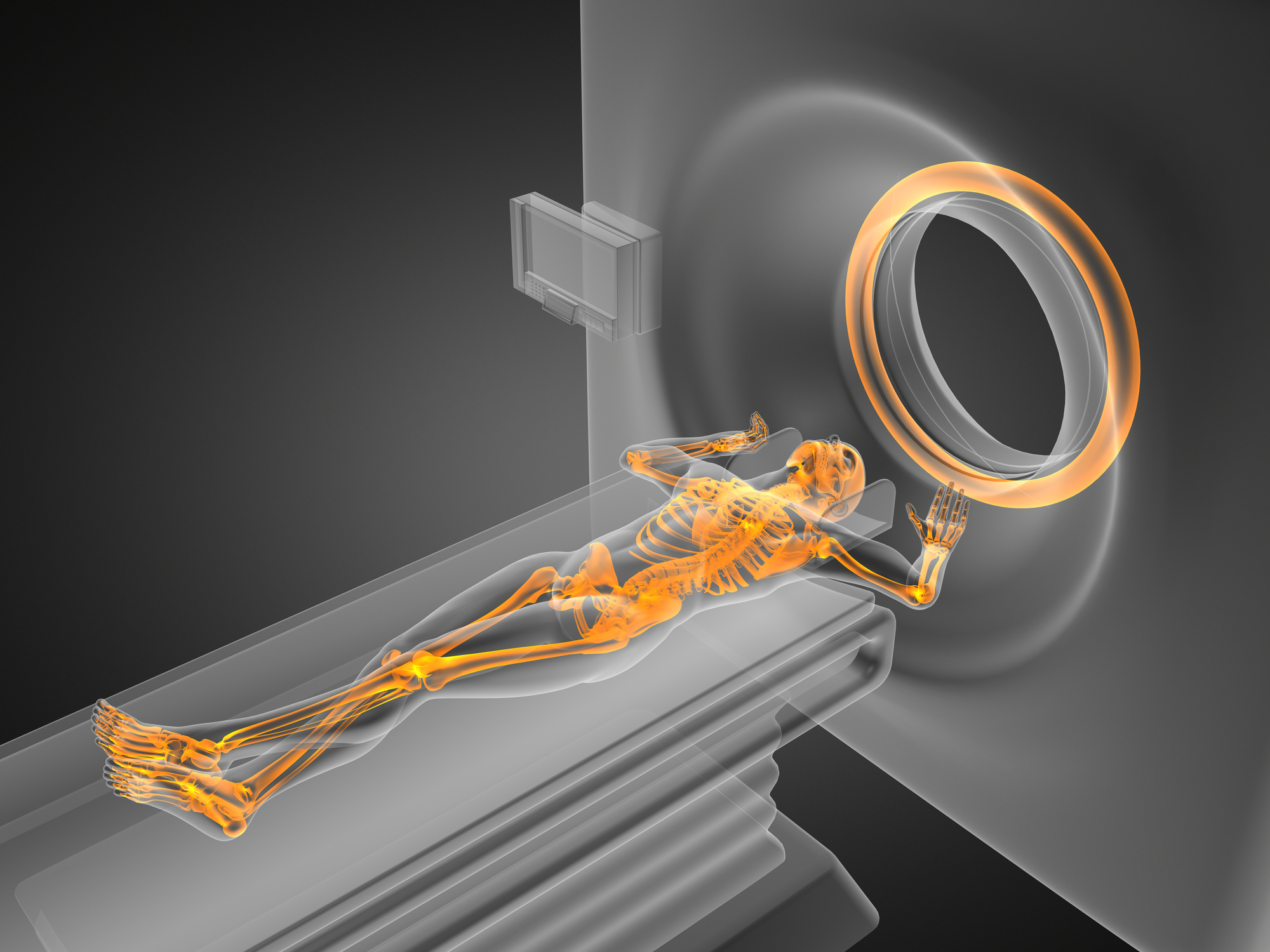
The development of 3D imaging techniques has taken these modalities to new horizons, offering healthcare professionals detailed, volumetric information that significantly enhances their ability to diagnose and treat various medical conditions. Creating a 3D image typically involves the reconstruction of multiple 2D slices or projections, which are then combined to form a comprehensive view of the body’s internal structures.
These 3D imaging technologies are employed in various medical applications. For example, they allow for more precise localisation and characterisation of tumours, improved diagnosis and management of cardiovascular diseases. In addition to enhanced surgical planning for orthopaedic procedures and better visualisation of the brain’s anatomy and function. Furthermore, 3D imaging has emerged as an invaluable tool for patient education, enabling healthcare professionals to communicate complex medical information to their patients and foster informed decision-making.
The future of 3D medical imaging is promising, as it is poised for numerous innovations and advancements in the near future. Integrating artificial intelligence (AI) and machine learning algorithms with 3D imaging is fascinating. This combination will transform medical imaging by automating image analysis, enhancing diagnostic accuracy, and personalising treatment planning. Based on ScienceSoft’s experience, CNN-powered analysis of 3D brain MRI scans enables automated tumour segmentation across multiple tissue types, achieving up to 87% accuracy and supporting neurologists in faster diagnosis, surgical planning, and treatment progress monitoring.
Furthermore, the fusion of 3D imaging with augmented reality (AR) and virtual reality (VR) technologies promises to revolutionise medical education, surgical planning, and patient engagement by offering immersive, interactive experiences.
The Evolution of 3D Medical Imaging
In recent years, advancements in medical imaging technology have continued to evolve, incorporating new techniques and applications that enhance the diagnostic capabilities of existing modalities. These major breakthroughs include:
- Digital radiography has replaced traditional film-based X-rays, offering improved image quality, faster processing times, and reduced radiation exposure for patients. In addition, digital images can be easily stored, shared, and analysed using specialised software.
- 3D ultrasound creates three-dimensional images of internal structures, while 4D ultrasound offers real-time 3D imaging, allowing physicians to observe the movement of organs and systems. This technology benefits obstetrics and fetal imaging, enabling clinicians to assess fetal development and identify abnormalities more effectively.
- Functional MRI measures changes in blood flow related to neural activity, allowing researchers and clinicians to map brain function and observe how different brain areas are involved in various tasks or stimuli. This technology has become essential in understanding brain disorders and planning brain surgeries.
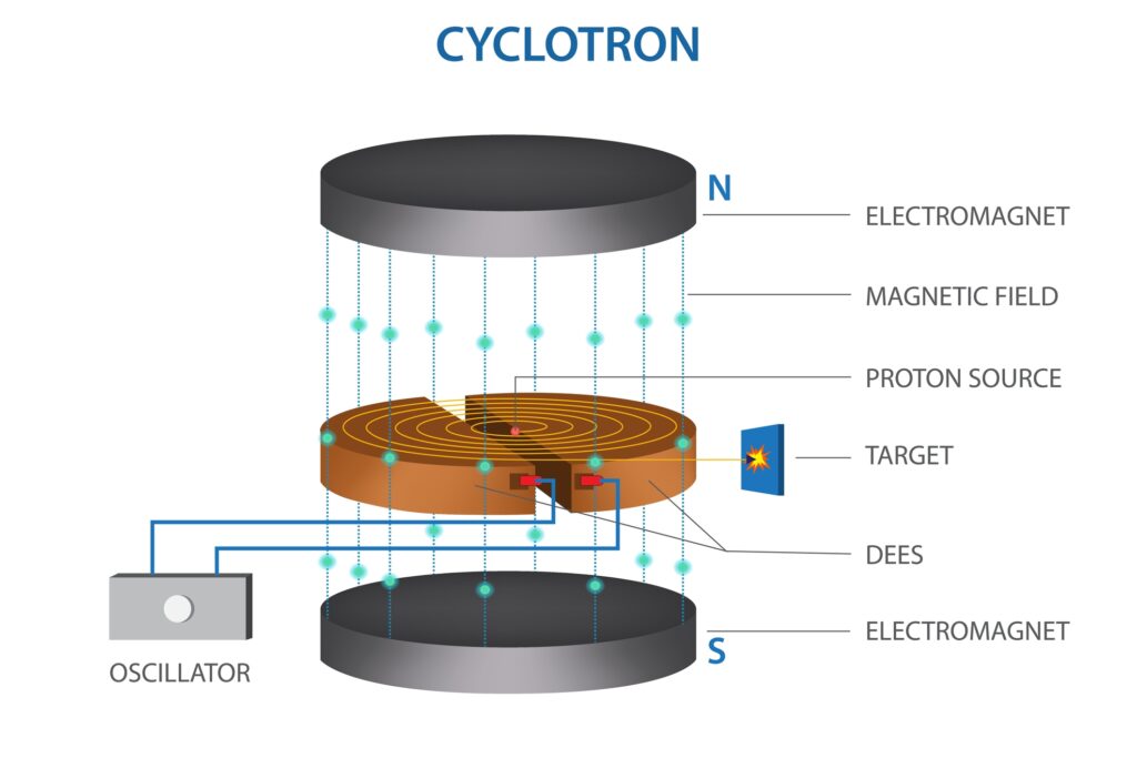
- Positron Emission Tomography (PET) scans utilise radioactive tracers to create three-dimensional images of the body’s internal processes, including metabolism and blood flow. PET scans are often used with CT or MRI scans to provide a more comprehensive view of a patient’s condition.
- Optical Coherence Tomography (OCT) is a non-invasive imaging technique that uses light to create high-resolution, cross-sectional images of biological tissues. It has become an essential tool in ophthalmology for diagnosing and monitoring various eye conditions, including age-related macular degeneration and diabetic retinopathy.
- AI and Machine Learning algorithms are increasingly integrated into medical imaging to improve image analysis, reduce the risk of human error, and increase efficiency. These technologies can help radiologists detect patterns and abnormalities more accurately and support decision-making in patient care.
- Image-Guided therapy, or interventional radiology, combines real-time imaging with minimally invasive procedures to diagnose and treat various conditions, including cancer, vascular diseases, and musculoskeletal disorders. Image-guided therapies often result in faster recovery and fewer complications than traditional surgical methods.
The Science Behind 3D Imaging
3D imaging involves the creation of a detailed 3D representation of an object, typically derived from a series of 2D images or data points. In medical imaging, 3D images are often reconstructed from multiple 2D slices or projections, providing clinicians with a comprehensive view of the body’s internal structures.
There are several techniques used to create 3D medical images, and these include:
- Volume Rendering is a technique that involves the conversion of 2D data (such as CT or MRI slices) into a 3D volume, with each voxel (3D pixel) assigned a specific colour and opacity based on its density or other properties. The final 3D image is then generated by compositing these voxels, allowing clinicians to visualise complex anatomical structures.
- Surface rendering involves extracting the surfaces of structures of interest from 2D data, creating a 3D mesh of the surface. This technique is particularly useful for visualising the shape and size of organs or other anatomical features.
- Multiplanar Reconstruction (MPR) involves the reformatting of 2D image data into different planes, allowing for the creation of 3D images that can be viewed from various angles.
Applications of 3D Imaging in Medicine
3D imaging in medicine has transformed how healthcare professionals diagnose, monitor, and treat various conditions, especially in oncology, cardiology, orthopaedics, neurology, dentistry, obstetrics and gynaecology.
The Impact of 3D Imaging on Cancer Care
Oncology, focused on the study and treatment of cancer, has witnessed a remarkable transformation in recent years. The most promising advancement is the integration of 3D imaging technology, which has emerged as a critical tool in the precise localisation and characterisation of tumours. This innovative approach is revolutionising how oncologists manage cancer care, allowing for more accurate staging, treatment planning, and monitoring of cancer progression or response to therapy.
3D imaging involves various techniques, such as CT, MRI, and PET, to generate detailed, multi-dimensional images of tumours within the body. These high-resolution images provide valuable insights into the tumours’ size, shape, and density, enabling clinicians to differentiate between benign and malignant lesions more effectively.
This level of precision is crucial for accurate cancer staging, which is essential for determining the best course of treatment. In addition, by understanding the extent and spread of cancer, oncologists can develop tailored treatment plans that take into account the unique characteristics of each patient’s tumour.
Furthermore, 3D imaging allows for more accurate treatment planning, particularly in the case of radiotherapy. By mapping the tumour’s exact location and surrounding healthy tissue, radiation oncologists can deliver targeted radiation doses, minimising damage to healthy tissues and reducing the risk of side effects.
Finally, 3D imaging is vital in monitoring cancer progression and response to therapy. As treatment progresses, these detailed images enable clinicians to evaluate the effectiveness of the therapy, detect any changes in tumour size, and identify potential recurrences at an early stage. This timely assessment allows for adjustments in treatment plans, ultimately improving patient outcomes and overall survival rates.
Unveiling the Heart’s Secrets with 3D Imaging Technology
Cardiology, the branch of medicine that deals with diagnosing and treating heart diseases, has been transformed by 3D imaging technology. This state-of-the-art innovation allows for a detailed examination of the heart’s structure and function, offering invaluable insights that aid in diagnosing and managing a wide range of cardiovascular diseases.
3D imaging techniques, such as echocardiography, cardiac MRI, and CT, provide high-resolution, real-time images of the heart, enabling clinicians to visualise its complex anatomy and assess its functionality. These images allow for a more accurate assessment of the heart’s size, shape, and contractile function and the identification of any abnormalities or defects within the heart’s valves and chambers.
One of the primary advantages of 3D imaging in cardiology is the ability to understand the heart’s function and blood flow. This is particularly crucial in diagnosing and managing conditions like valvular heart disease, congenital heart defects, and cardiomyopathies. In addition, by providing a precise evaluation of these conditions, 3D imaging assists in determining the most appropriate course of treatment and monitoring the patient’s response to therapy.
Furthermore, 3D imaging plays a significant role in the planning and execution of minimally invasive cardiac procedures, such as transcatheter aortic valve replacement (TAVR) and transcatheter mitral valve repair (TMVR). By facilitating accurate measurements and visualisations, these advanced imaging techniques help to reduce the risk of complications during these procedures, ultimately improving patient outcomes.
In addition to diagnosis and treatment planning, 3D imaging is invaluable for monitoring patients with cardiovascular diseases. Regular assessments enable clinicians to track disease progression, evaluate the effectiveness of prescribed treatments, and make any necessary adjustments to the treatment plan, ensuring optimal patient care.
Orthopaedic Breakthroughs: Enhancing Patient Care with 3D Imaging Technology
Orthopaedics, the branch of medicine concerned with diagnosing, treating, and preventing musculoskeletal disorders, has greatly benefitted from advancements in 3D imaging technology. This cutting-edge innovation transforms the planning and execution of orthopaedic surgeries, including joint replacement, spinal fusion, and fracture repair, ultimately improving patient outcomes and overall quality of care.
3D imaging techniques, such as CT and MRI, generate detailed images of bones, joints, and soft tissues, providing invaluable insights for orthopaedic surgeons. These high-resolution images enable a comprehensive understanding of the patient’s anatomy, allowing for precise surgical planning and minimising potential complications.
In joint replacement surgeries, such as hip and knee arthroplasty, 3D imaging facilitates accurate measurements of joint components and customisation of prosthetic implants. By ensuring optimal sizing and alignment, surgeons can improve the longevity of the implant, reduce the risk of complications, and enhance postoperative function and patient satisfaction.
Spinal fusion surgeries also benefit from 3D imaging technology. The detailed visualisation of the spine’s anatomy, including the vertebrae, discs, and surrounding soft tissues, enables surgeons to make more informed decisions regarding the optimal approach and techniques for spinal stabilisation. This level of precision contributes to a higher success rate and a reduced risk of complications, such as nerve damage or spinal instability.
In fracture repair, 3D imaging assists in identifying complex fractures and assessing surrounding soft tissue damage. This information is critical in determining the most appropriate method of fracture fixation, whether internal or external. Furthermore, 3D imaging helps to guide the surgical procedure, ensuring proper alignment and stabilisation of the fractured bone and promoting successful healing and recovery.
Neurology Transformed by 3D Imaging Technology
Neurology is focused on diagnosing and treating neurological disorders and has been significantly advanced by integrating 3D imaging technology. This groundbreaking innovation offers detailed views of the brain’s anatomy and function, playing a crucial role in diagnosing and treating various neurological disorders, including brain tumours, aneurysms, and neurodegenerative diseases.
3D imaging techniques, such as MRI, CT, and PET, generate high-resolution images of the brain’s structure and function. These detailed images enable neurologists and neurosurgeons to identify and assess abnormalities, such as tumours or vascular malformations, with unprecedented precision.
In the case of brain tumours, 3D imaging provides essential information regarding the size, location, and extent of the lesion. This information is critical for accurate diagnosis, staging, and treatment planning, whether it involves surgery, radiation therapy, or chemotherapy. Moreover, 3D imaging assists in differentiating between benign and malignant tumours and monitors treatment response and potential recurrence.
For patients with cerebral aneurysms, 3D imaging offers crucial insights into the size, shape, and location, enabling clinicians to determine the risk of rupture and the most appropriate treatment approach, such as endovascular coiling or surgical clipping. By providing a comprehensive understanding of the aneurysm’s characteristics, 3D imaging helps to reduce the risk of complications and improve patient outcomes.
In neurodegenerative diseases like Alzheimer’s, Parkinson’s, and multiple sclerosis, 3D imaging contributes to a greater understanding of the disease’s underlying pathological processes and progression. These insights facilitate early diagnosis and intervention and the development of targeted therapies that may slow or halt disease progression.
The Role of 3D Ultrasound in Obstetrics and Gynaecology
Obstetrics and Gynaecology, the branch of medicine focusing on the female reproductive system and prenatal care, has experienced significant advancements with the introduction of 3D ultrasound imaging technology. This innovative technology enables clinicians to visualise the developing fetus, assess fetal health, and detect abnormalities in the placenta, uterus, and other pelvic structures, improving prenatal care and patient outcomes.
3D ultrasound imaging is a non-invasive diagnostic tool that generates detailed, real-time images of the fetus and its surroundings using sound waves. Unlike traditional 2D ultrasound, 3D ultrasound offers a more comprehensive view of the fetus, providing information about its anatomy, growth, and overall health. This technique also allows for better visualisation of the placenta, umbilical cord, and amniotic fluid, which is essential for fetal well-being.
One of the primary benefits of 3D ultrasound in obstetrics and gynaecology is the early detection of congenital abnormalities, such as cleft lip, heart defects, and skeletal anomalies. By identifying these issues in the prenatal stage, clinicians can provide appropriate counselling, plan for necessary interventions, and improve the overall prognosis for the affected infants.
In addition to its use in prenatal care, 3D ultrasound imaging is valuable in diagnosing and managing gynaecological conditions, such as uterine fibroids, endometriosis, and ovarian cysts. By offering a detailed view of the uterus, ovaries, and other pelvic structures, this technology aids in determining the most effective course of treatment and monitoring response to therapy.
Moreover, 3D ultrasound is useful for assessing the position and presentation of the fetus in the later stages of pregnancy, guiding decisions regarding the mode of delivery and reducing the risk of complications during labour and birth.
The Impact of 3D Imaging on Modern Dentistry
Dentistry, the branch of medicine that focuses on diagnosing, treating, and preventing oral health conditions, has greatly benefitted from introducing 3D imaging technologies. Cone-beam computed tomography (CBCT) is one such technology that provides comprehensive information about the teeth, jaws, and surrounding structures, significantly improving treatment planning for dental procedures and oral surgery.
CBCT is a specialised form of X-ray imaging that generates high-resolution, 3D images of the teeth, jaws, and associated structures with minimal radiation exposure. This advanced imaging technique offers a more comprehensive view of the patient’s oral anatomy than traditional 2D X-rays, allowing for enhanced diagnostic accuracy and precision in treatment planning.
One of the primary applications of CBCT in dentistry is in the planning and execution of dental implant procedures. By providing detailed information about bone density, quality, and available space for implant placement, CBCT enables dentists to ensure optimal implant positioning and avoid potential complications, such as nerve or sinus damage.
Additionally, 3D imaging is invaluable in diagnosing and treating orthodontic issues, such as malocclusion and impacted teeth. CBCT allows orthodontists to assess tooth and jaw alignment, plan for braces or clear aligner therapy, and monitor the progress of orthodontic treatment more effectively.
3D imaging is also essential in oral and maxillofacial surgery, including corrective jaw surgery, wisdom tooth extraction, and treating temporomandibular joint (TMJ) disorders. CBCT provides critical insights into the patient’s anatomy, enabling surgeons to minimise the risk of complications and optimise surgical outcomes.
Furthermore, CBCT aids in identifying and managing dental pathologies, such as cysts, tumours, and infections, by offering detailed images of the affected areas and assisting in developing targeted treatment plans.
Shaping Aesthetics: The Role of 3D Imaging in Plastic Surgery
Plastic surgery has been transformed by the integration of 3D imaging technology, which allows surgeons to plan procedures with greater precision and provide patients with realistic previews of their potential results.
In breast procedures such as augmentation, reduction, and reconstruction, 3D imaging enables surgeons to visualise changes directly on a virtual model of the patient’s body. The Breast Reduction Surgeons of Long Island, whose specialty is breast reduction in Long Island, note that this technology helps patients preview size and shape adjustments, making the decision process more transparent and reassuring.
For facial procedures like rhinoplasty or facelifts, 3D imaging also provides detailed mapping of bone structure and soft tissue, helping surgeons design more balanced, natural-looking results. This added precision not only improves outcomes but also enhances patient confidence going into surgery.
Advantages of 3D Imaging in Medicine
3D images provide a more accurate and comprehensive view of internal structures, allowing healthcare professionals to understand complex anatomical relationships better and identify abnormalities that may not be apparent in 2D images.
By offering a complete picture of the body’s internal structures, 3D imaging can improve diagnostic accuracy, leading to more targeted and effective treatments.
3D images can be used to create detailed surgical plans, reducing the risk of complications and improving surgical outcomes. In some cases, 3D images can also be used during surgery to provide real-time guidance and ensure the accurate placement of surgical instruments.
3D images can help patients better understand their medical condition and the proposed treatment plan, increasing patient engagement and satisfaction.
Limitations and Challenges of 3D Imaging in Medicine
Although its numerous advantages, 3D medical imaging is not without its limitations and challenges:
The generation and manipulation of 3D images require substantial computing power and specialised software, which can be expensive and time-consuming.
Some 3D imaging techniques, such as CT scans, involve exposure to ionising radiation. While efforts are continuously being made to minimise radiation doses, the risk of radiation exposure must always be considered, particularly in pregnant women and children.
The quality of 3D images can be affected by various factors, including patient movement, image noise, and artefacts. These issues can potentially impact the diagnostic accuracy and utility of 3D imaging.
The interpretation of 3D images can be more complex than traditional 2D images, requiring additional training and expertise from healthcare professionals.
Future Trends in 3D Imaging in Medicine
In the coming years, integrating AI and machine learning algorithms is expected to revolutionise 3D imaging in medicine. These advanced technologies have the potential to significantly enhance the speed and accuracy of image processing, segmentation, and analysis. This will allow healthcare professionals to quickly identify abnormalities and make informed decisions regarding diagnosis and treatment. Furthermore, AI-driven algorithms can be trained to recognise patterns in complex datasets, facilitating early detection of diseases and improving patient outcomes.
Advancements in visualisation techniques, such as holography and virtual reality (VR), are also expected to reshape how medical professionals interact with 3D images. Holography enables the creation of 3D images without special glasses, while VR allows for an immersive, interactive experience in a simulated environment. These technologies can provide healthcare practitioners with more intuitive and effective ways to explore and manipulate 3D medical images, enhancing their understanding of the underlying anatomy and pathology.
Another emerging trend in 3D imaging is the fusion of multiple imaging modalities, such as PET/CT or PET/MRI, to provide more comprehensive and accurate diagnostic information. By combining the strengths of different imaging techniques, healthcare professionals can better understand a patient’s condition, particularly in complex cases or when multiple physiological processes need to be assessed simultaneously. This can lead to more accurate diagnoses and tailored treatment plans, ultimately improving patient care and outcomes.
The development of personalised medicine is another area where 3D imaging is poised to make a significant impact. 3D imaging can help tailor treatments to each patient’s needs by offering detailed insights into an individual’s unique anatomy and physiology. This personalised approach can lead to more effective therapies, reduced side effects, and improved patient satisfaction.
Finally, advancements in 3D imaging technology are expected to enhance the field of telemedicine, enabling remote consultations and collaboration between healthcare professionals across the globe. Specialists in different locations can share and analyse high-quality, real-time 3D images, facilitating expert opinions and improving patient care, even in remote or underserved areas.
Disclaimer
The content provided in this article, A Dive into the World of 3D Medical Imaging, is for informational and educational purposes only and is not intended to serve as medical advice, diagnosis, or treatment. While every effort has been made to ensure the accuracy of the information at the time of publication, Open Medscience makes no representations or warranties, express or implied, about the completeness, accuracy, reliability, suitability, or availability of the content.
The technologies, techniques, and clinical applications discussed are continually evolving. Readers are advised to consult with qualified healthcare professionals or medical specialists for advice tailored to their individual circumstances. Open Medscience does not endorse any specific product, service, or technology mentioned in this article.
Any views or opinions expressed are those of the authors and do not necessarily reflect the official policy or position of Open Medscience or its affiliates.
Use of this article and any reliance on the information contained herein is at the reader’s own risk.