Cell-based assays are becoming increasingly important in modern biology and drug discovery. They provide context that biochemical assays alone cannot offer, capturing the complexity of living systems and giving researchers deeper insights into cellular behaviour. Advances in detection technology now make it possible to integrate cell imaging workflows into high-throughput plate formats. A dedicated microplate fluorometer is at the centre of this shift, enabling sensitive, reproducible measurements for assays ranging from viability studies to reporter gene expression.
Why Fluorescence Matters for Cell Imaging
Fluorescence detection allows researchers to track dynamic processes in real time, including protein expression, intracellular signalling, and cell proliferation. Unlike absorbance-based methods, fluorescence offers higher sensitivity and the ability to multiplex multiple readouts from the same well. This makes it indispensable for imaging applications where subtle biological changes need to be detected quickly and accurately.
Applications of Cell Imaging in Plate-Based Workflows
- Cell Viability and Cytotoxicity: Fluorescent dyes and reporters measure live/dead ratios, mitochondrial activity, or membrane integrity, providing key insights into drug safety and efficacy.
- Reporter Gene Assays: Fluorescent proteins such as GFP or RFP allow researchers to study gene regulation and signalling pathways under controlled experimental conditions.
- Intracellular Ion Tracking: Calcium and pH-sensitive dyes are commonly used to monitor cell signalling, a critical component of pharmacological screening.
- Apoptosis and Cell Cycle Analysis: Specialised fluorescence probes track programmed cell death or cell cycle progression, offering a detailed view of cellular responses.
The Role of Microplate Fluorometers
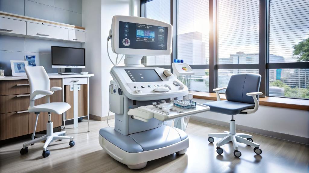
Plate-based fluorometers combine sensitive optics with high-throughput capabilities, supporting cell imaging in 96- to 1536-well formats. Features such as adjustable gain, temperature regulation, and kinetic reading modes allow them to capture both endpoint and time-lapse data. Unlike microscopy, which can be labour-intensive, fluorometers enable scalable imaging for larger sample sets, making them especially valuable for screening applications.
Advantages of Plate-Based Cell Imaging
- High Throughput: Hundreds of samples can be measured simultaneously, dramatically increasing efficiency.
- Consistency: Automated plate handling ensures uniform conditions across wells.
- Scalability: Researchers can move seamlessly from small pilot assays to large compound libraries.
- Integration: Compatible with automation and analysis software, fluorometers fit easily into existing lab workflows.
Conclusion
Cell imaging has evolved from a specialised, microscopy-only approach to a scalable, plate-based method that meets the demands of modern research. A microplate fluorometer provides the sensitivity, flexibility, and throughput needed to capture detailed cellular information at scale. For labs seeking to combine the biological relevance of live-cell assays with the efficiency of high-throughput workflows, plate-based fluorometric imaging is a critical innovation.
Frequently Asked Questions
What makes fluorescence suitable for cell imaging?
Fluorescence offers high sensitivity, multiplexing capabilities, and real-time tracking of cellular processes, making it ideal for imaging assays.
How does a microplate fluorometer differ from a microscope?
While microscopes provide detailed images of individual cells, fluorometers measure bulk fluorescence across many wells, allowing for high-throughput quantification.
Can cell imaging assays be scaled for drug discovery?
Yes. Plate-based formats supported by fluorometers enable rapid screening of large compound libraries while maintaining biological relevance.
What types of cell-based assays benefit most from fluorometry?
Viability, apoptosis, reporter gene expression, intracellular ion tracking, and cytotoxicity assays are all well-suited to fluorometric detection.
Is specialised software needed for cell imaging assays?
Yes. Analysis software supports kinetic measurements, normalisation, and data visualisation, ensuring reliable interpretation of complex results.
Disclaimer
The information provided in this article, Cell Imaging with Microplate Fluorometers: Expanding the Boundaries of Assay Design, is intended for educational and informational purposes only. It does not constitute professional advice, diagnosis, or endorsement of any specific product, technology, or methodology. While efforts have been made to ensure the accuracy and relevance of the content at the time of publication, Open MedScience makes no representations or warranties, express or implied, regarding its completeness, reliability, or suitability for any particular purpose. Readers are encouraged to verify any technical details, follow appropriate laboratory safety procedures, and consult qualified professionals before applying any techniques or protocols discussed. Open MedScience shall not be liable for any loss, injury, or damage arising from the use or reliance on the information contained herein.


