
Fluorine-18: A Cornerstone Radionuclide in Modern Medical Imaging
 By
By
Fluorine-18 Medical Imaging revolutionises diagnostic techniques by enabling precise visualisation of metabolic processes in the body.
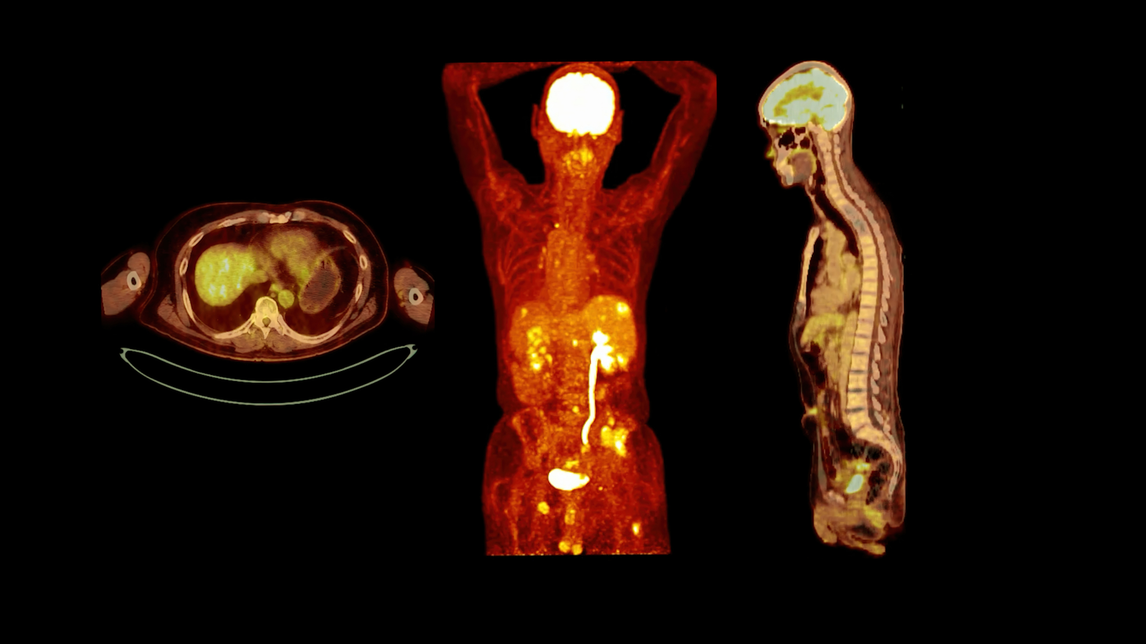
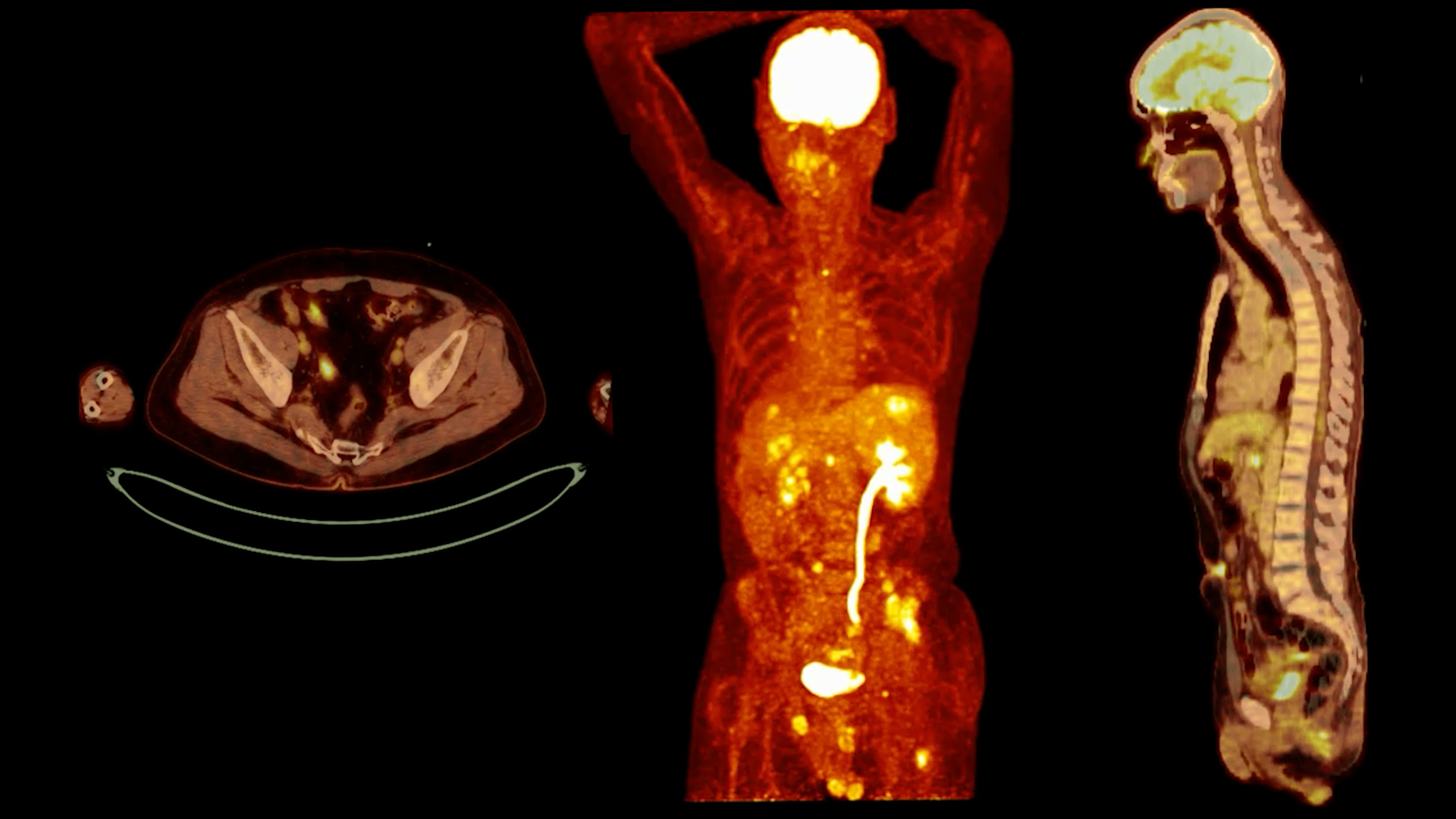
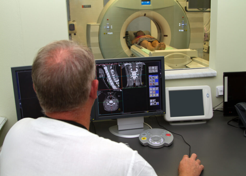
A positron emission tomography (PET) scan is a cutting-edge medical imaging technique that visualises and measures the metabolic activity of the body’s tissues and organs. By providing detailed information on the cellular level, positron emission tomography scans have revolutionised the diagnosis, staging, and treatment of numerous diseases, including cancer, neurological disorders, and cardiovascular diseases.
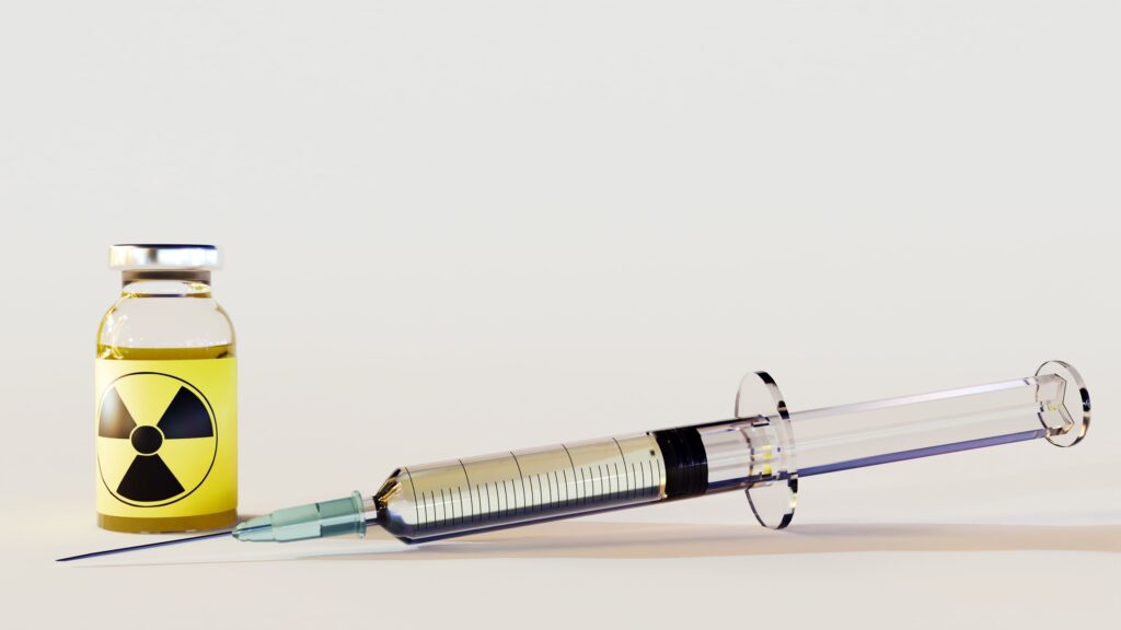
These scans rely on detecting positrons, subatomic particles emitted during the radioactive decay of certain isotopes. The procedure involves the injection of a radiotracer, a compound that includes a small amount of a radioactive substance called a radionuclide. The most commonly used radiotracer is fluorodeoxyglucose (FDG), a molecule similar to glucose. FDG is absorbed by cells throughout the body, with cancerous and other highly active cells taking up more of the compound than normal cells.
As the radionuclide decays, it emits positrons that collide with nearby electrons, resulting in the annihilation of both particles and the production of two gamma-ray photons. These photons travel in opposite directions and are detected by a ring of detectors surrounding the patient. By analysing the timing and location of these detections, a computer can generate a three-dimensional image of the body’s tissues, indicating areas of high and low metabolic activity.
These scans offer advantages over other imaging techniques, such as computed tomography (CT) and magnetic resonance imaging (MRI). The ability to measure metabolic activity allows for early disease detection and accurate assessment of treatment response. Moreover, these scans can be combined with CT or MRI to provide detailed anatomical and functional information in a single study.
Despite its many benefits, PET scans have limitations. Radioactive tracers may raise concerns about radiation exposure, although the risks are generally low. Additionally, these scans can be costly and not covered by all insurance plans. Finally, PET scans may yield false-positive or false-negative results, particularly in cases of inflammation or infection, which can also demonstrate increased metabolic activity.
home »
By
Fluorine-18 Medical Imaging revolutionises diagnostic techniques by enabling precise visualisation of metabolic processes in the body.
 By
By
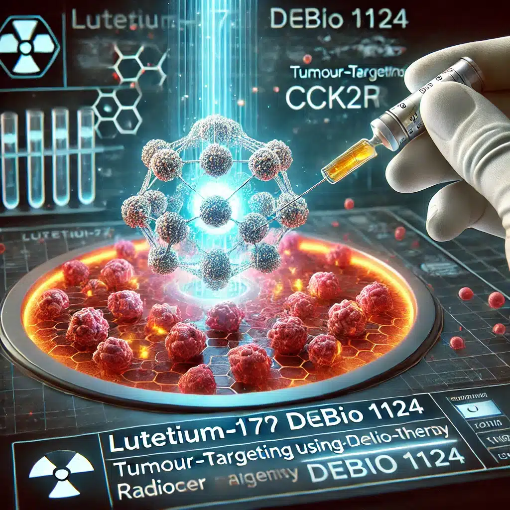
Lutetium-177 Debio 1124, a second-generation theranostic agent, selectively targets CCK2R-expressing tumours, offering precision radiotherapy and personalised oncology advancements.
 By
By
Green radiology promotes sustainability by reducing energy use, managing waste, and integrating renewable resources effectively.
 By
By
Advances in medical imaging technology have significantly improved diagnostic accuracy, enabling earlier detection and more personalised treatments.
 By
By
Diagnostic imaging in motor neurone disease (MND) is crucial for early detection, disease monitoring, and differentiating from other conditions.
 By
By
Radiopharmaceuticals in diagnostics provide critical insights into disease processes, improving accuracy, patient care, and treatment outcomes significantly.
 By
By
PET scans are crucial for detecting metabolic activity, providing valuable insights into cancer, neurological disorders, and cardiovascular diseases.
 By
By
Fluorine-18 Fluoroestradiol PET imaging enables early breast cancer detection, accurate staging, and treatment response monitoring, improving patient outcomes.
 By
By
PET imaging (Positron Emission Tomography) is an established diagnostic imaging tool used in Nuclear Medicine.