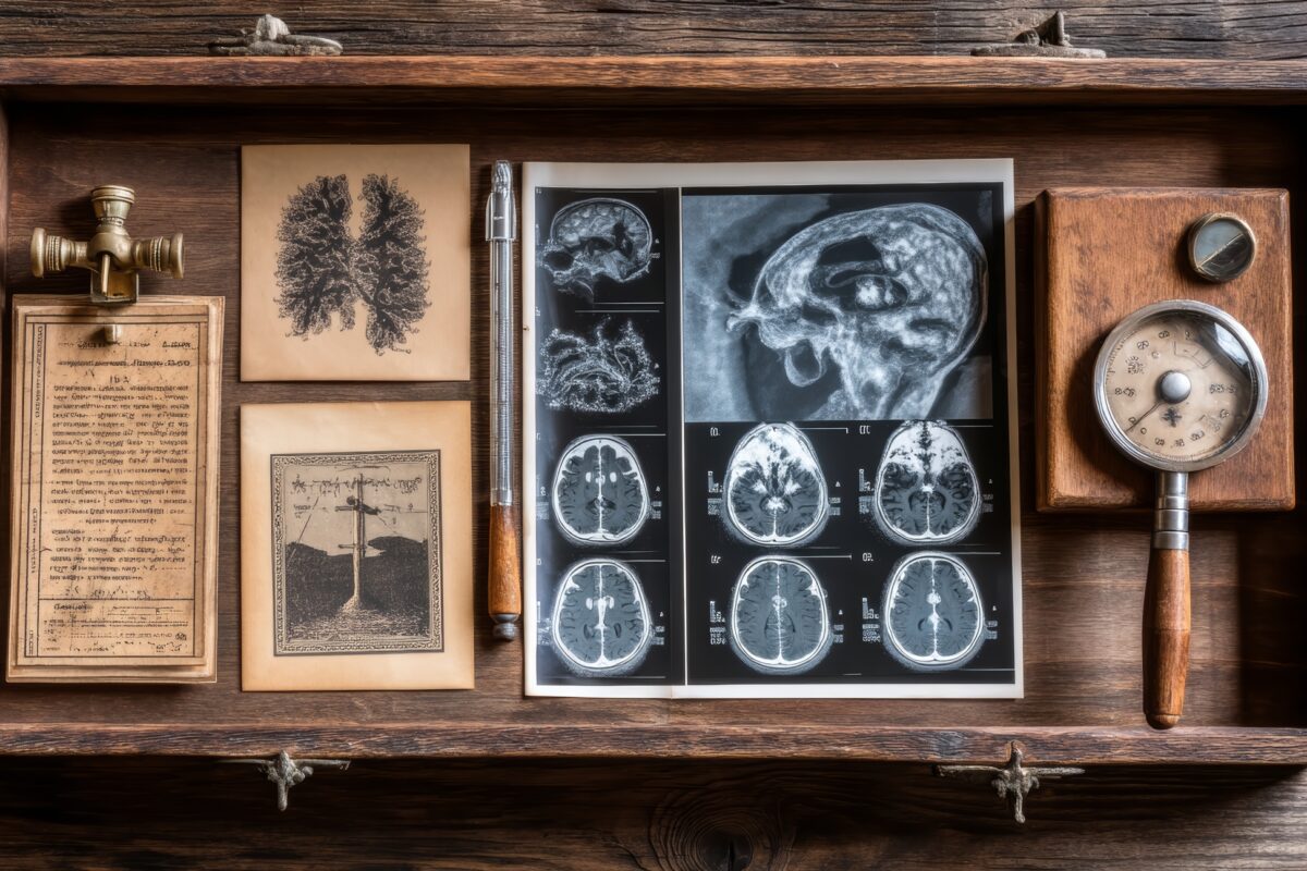
The Pioneers of Medical Imaging: The People Who Changed the Way We See Ourselves
 By
By
Explore the groundbreaking work of the pioneers of medical imaging and how they changed the landscape of modern medicine.
By
Explore the groundbreaking work of the pioneers of medical imaging and how they changed the landscape of modern medicine.
 By
By
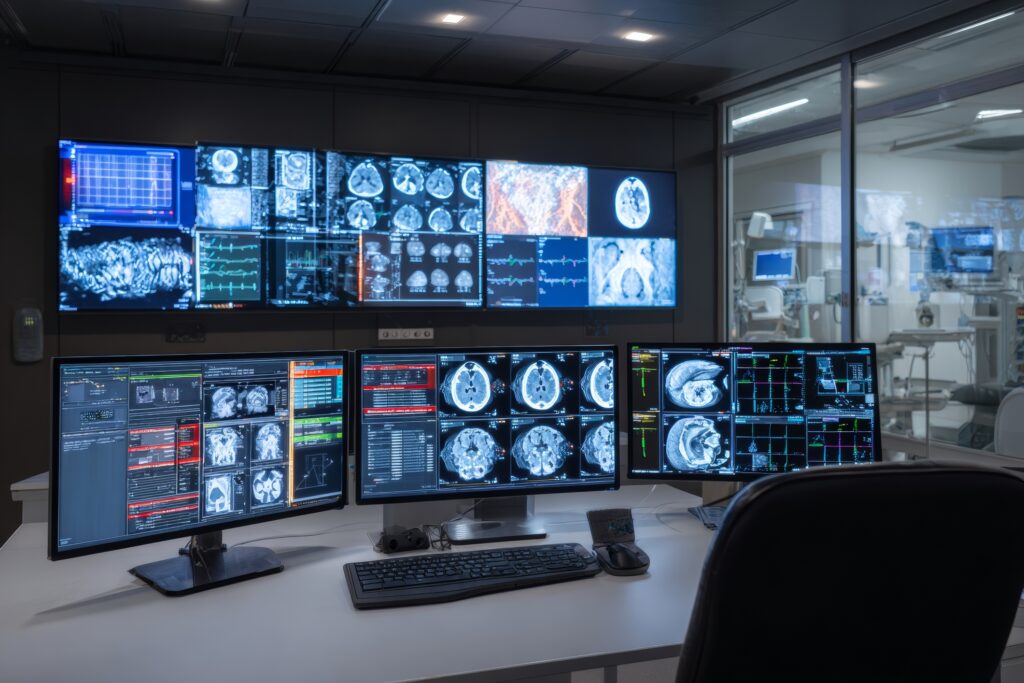
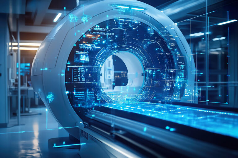
Discover the fascinating types of medical imaging, from X-rays to advanced molecular techniques, shaping health care today.
 By
By
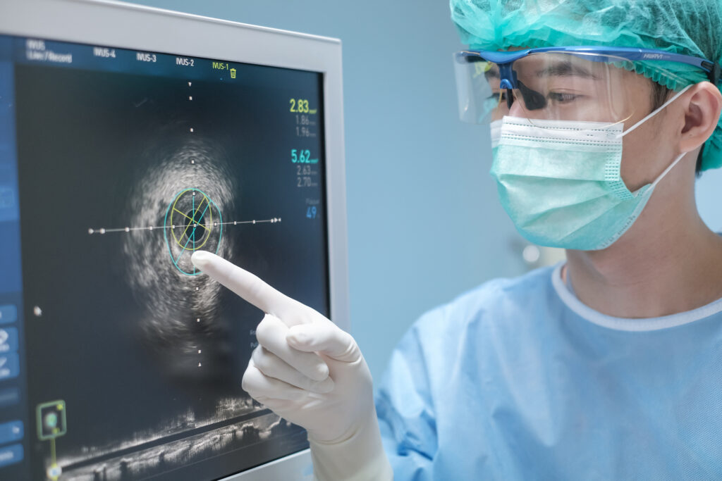
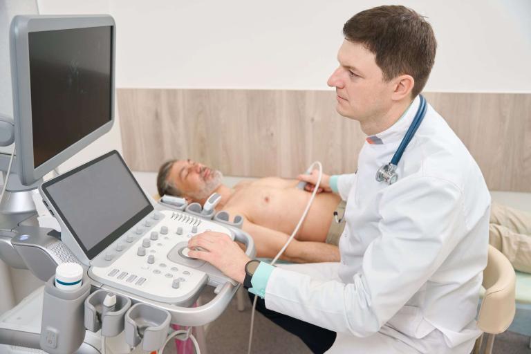
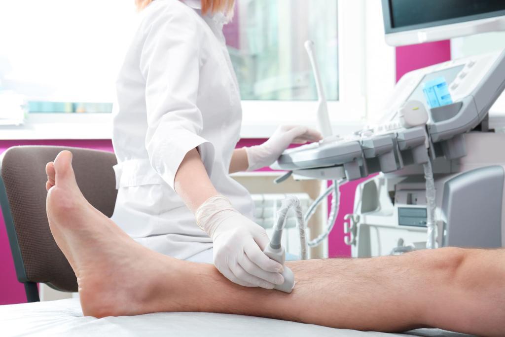
Discover how ultrasound imaging in healthcare provides accurate information. Learn its use in obstetrics, cardiology, and more. Image for illustration only. Person depicted is a model.
 By
By
Evaluate your expertise with the medical imaging quiz, focusing on imaging parameters, radiation safety, and practical case studies.
 By
By
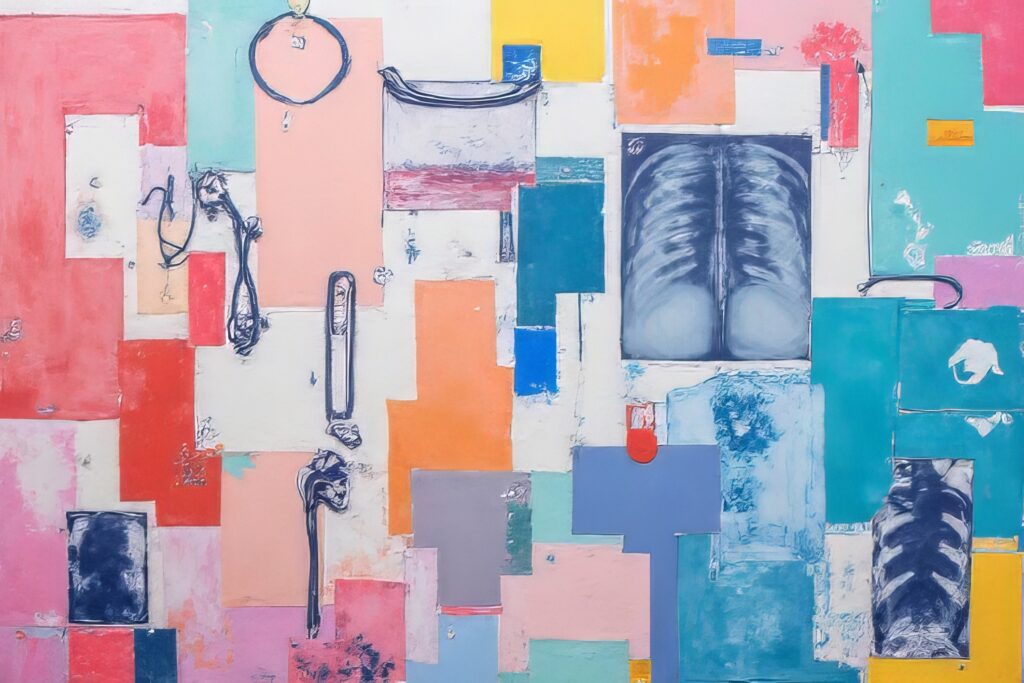
Uncover the truth about castor oil and medical imaging. Learn why its benefits remain anecdotal and unverified by science.
 By
By
Learn about the UK Biobank’s pioneering imaging study that has delivered unique insights from 100,000 participant scans.
 By
By
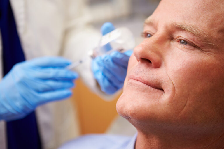
Cost of Botox injections may increase when medical imaging, like ultrasound, is used to guide accurate placement. Image for illustration only. People depicted are models.
 By
By
The rise of online nursing schools empowers aspiring nurses through flexible, accessible, and affordable educational opportunities worldwide. Image for illustration only. Person depicted is a model.
 By
By
New Year medical imaging breakthroughs highlight innovative technologies, transforming healthcare diagnostics and improving patient outcomes worldwide.
 By
By
Ultrasound scanning provides a non-invasive way to visualise organs, detect abnormalities, and assess soft tissue health. Image for illustration only. People depicted are models.
 By
By
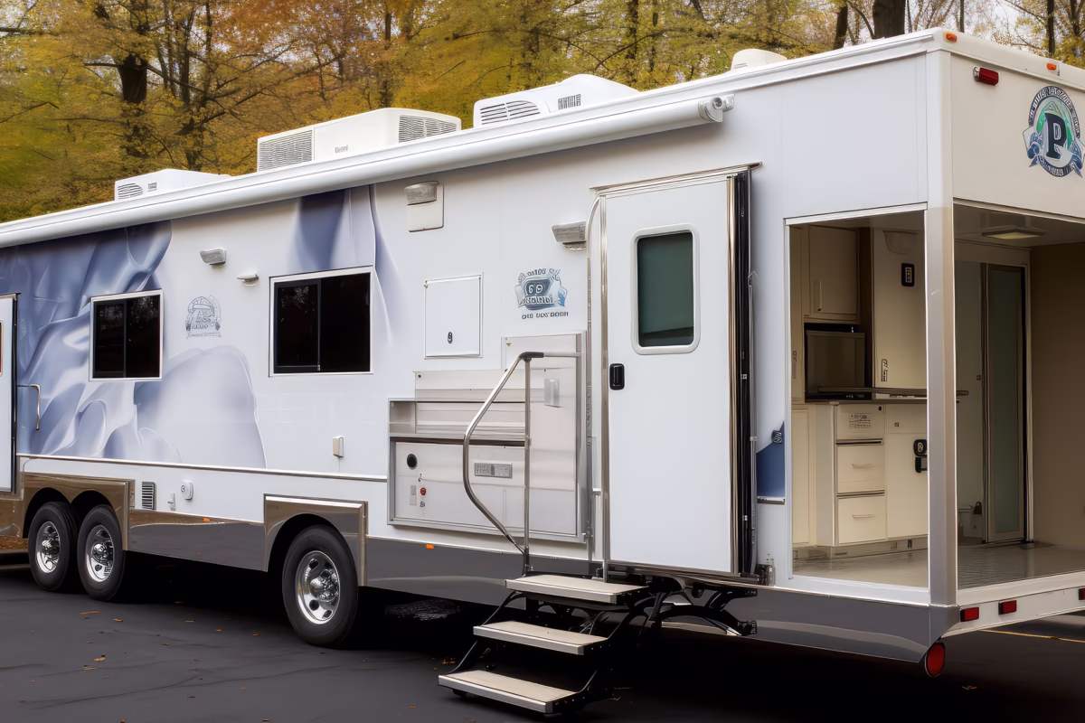
Medical mobile scanners revolutionise healthcare by providing portable, accessible imaging solutions for remote, emergency, and underserved areas.
 By
By
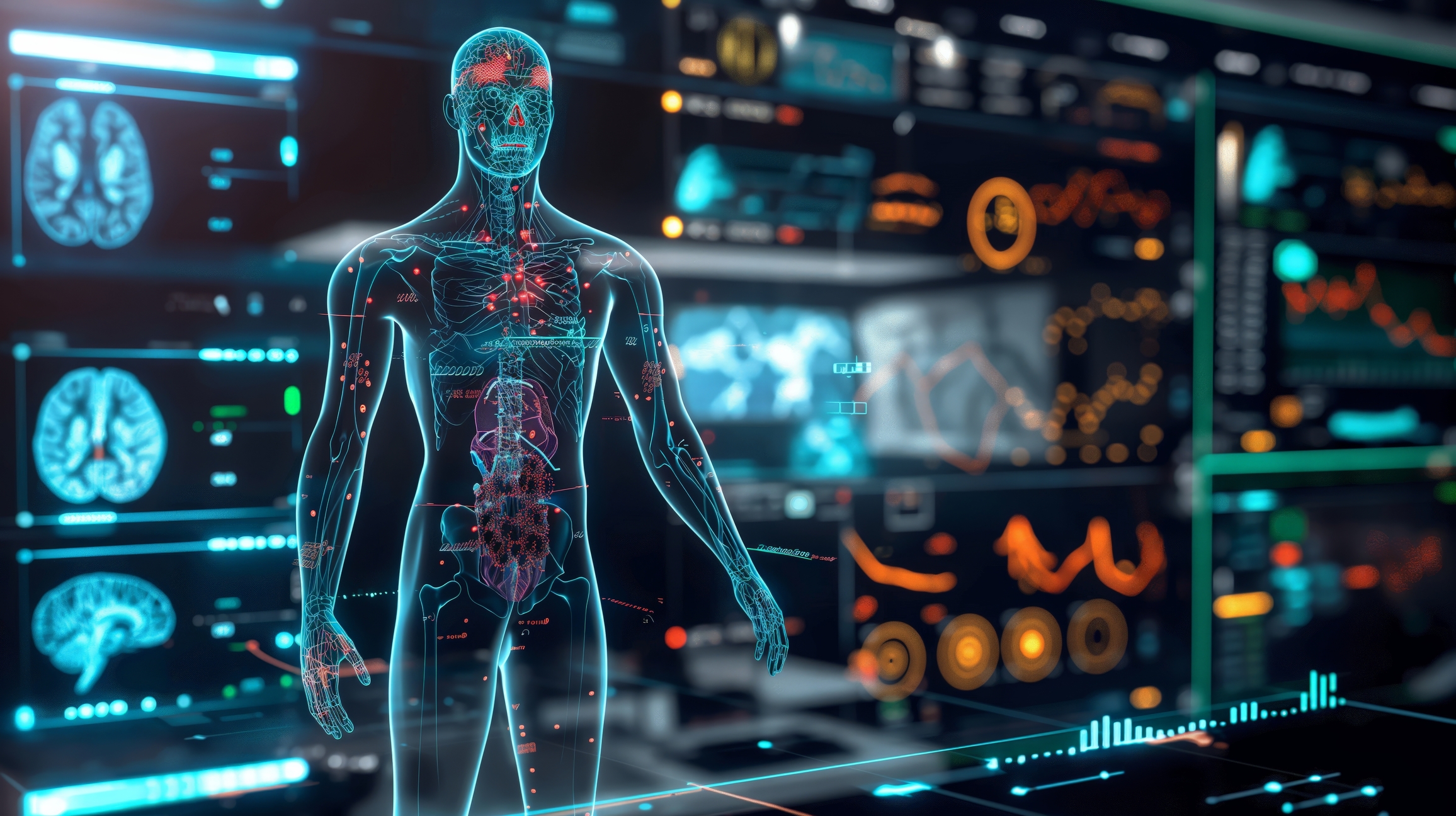
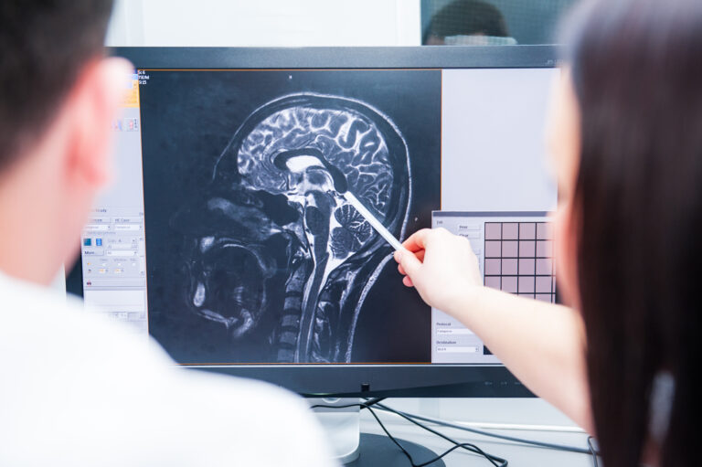
Advances in medical imaging technology have significantly improved diagnostic accuracy, enabling earlier detection and more personalised treatments.
 By
By
The article discusses the integration of technology in women’s health, offering advanced, accessible solutions for enhanced well-being and medical care.
 By
By
Diagnostic ultrasound imaging, a non-invasive technique, safely visualises internal organs, aiding in diverse medical diagnoses effectively.
 By
By
To excel in medical imaging, one must pursue postgraduate education beyond basic nursing or medicine. Image for illustration only. People depicted are models.
 By
By
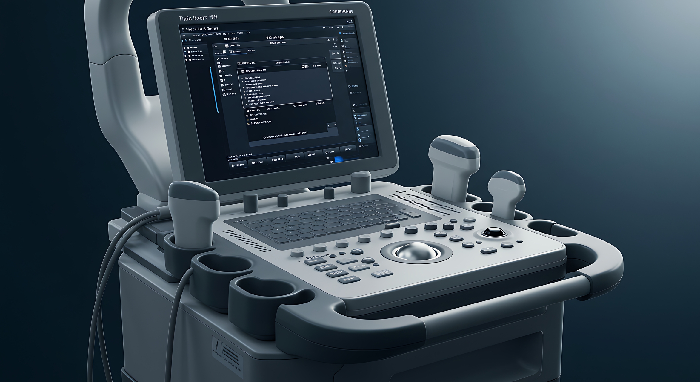
Point of Care imaging’s evolution, marked by miniaturisation, has revolutionised bedside diagnostics and patient care delivery.
 By
By
Photoacoustic imaging fuses optical specificity with ultrasound depth, revolutionising deep-tissue visualization in biomedical diagnostics and research.
 By
By
Micro-ultrasound’s detailed imaging necessitates expert analysis, mindful safety practices, and promises future multimodal diagnostic integrations.
 By
By
EMAI combines electromagnetic fields with ultrasound to enhance non-destructive testing and medical diagnostics with high-resolution imaging.
 By
By
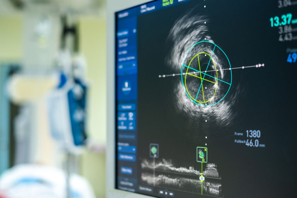
Ultrasound Localization Microscopy offers unprecedented microvascular insights, transforming diagnosis and treatment through super-resolution imaging.
 By
By
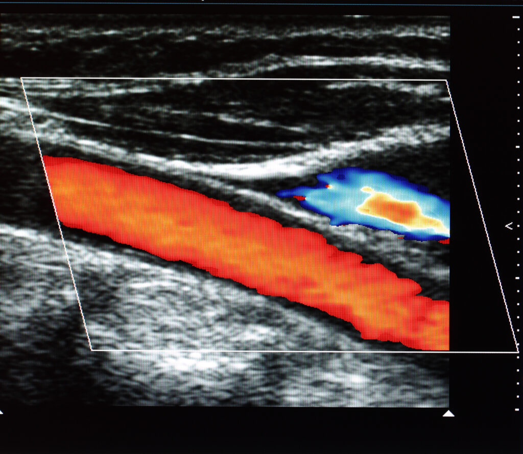
Ultrafast Doppler imaging offers unprecedented real-time insights into blood flow for improved diagnostic accuracy in medicine.
 By
By
Superb Microvascular Imaging transforms diagnostics, revealing minute vessels with clarity, sans contrast agents, enhancing patient care.
 By
By
Magnetomotive Ultrasound combines magnetic nanoparticles with ultrasound imaging for enhanced disease diagnosis and targeted therapeutic interventions. Image for illustration only. People depicted are models.
 By
By
Topics on PET imaging, automated radiosynthesis, breast cancer, prostate cancer therapy and ultrasound.