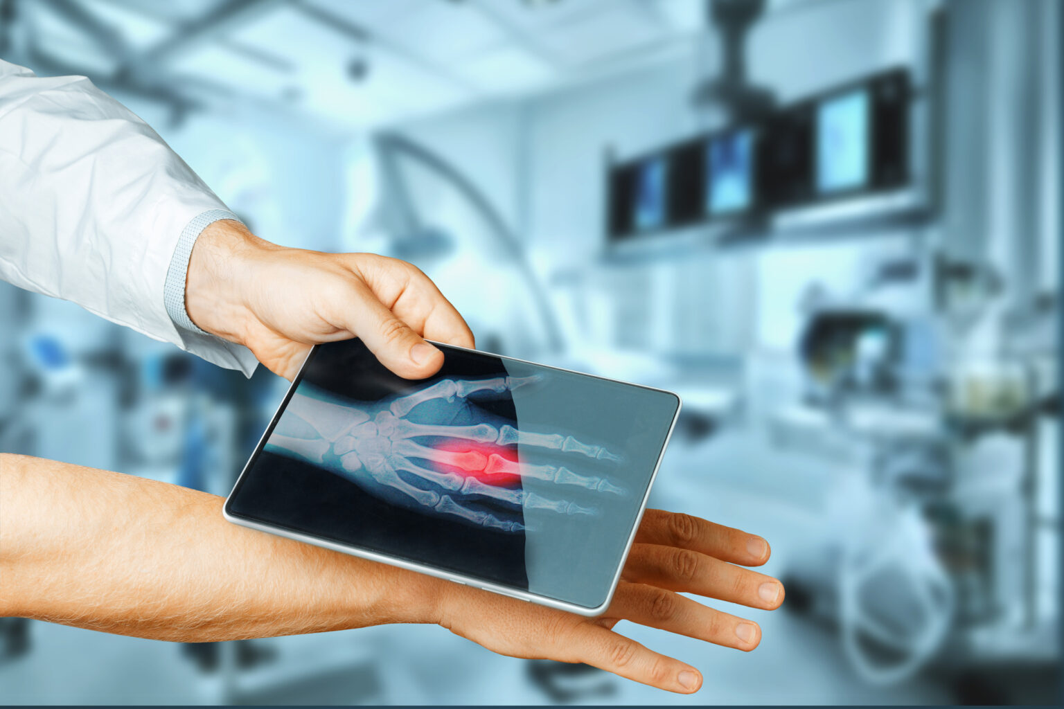
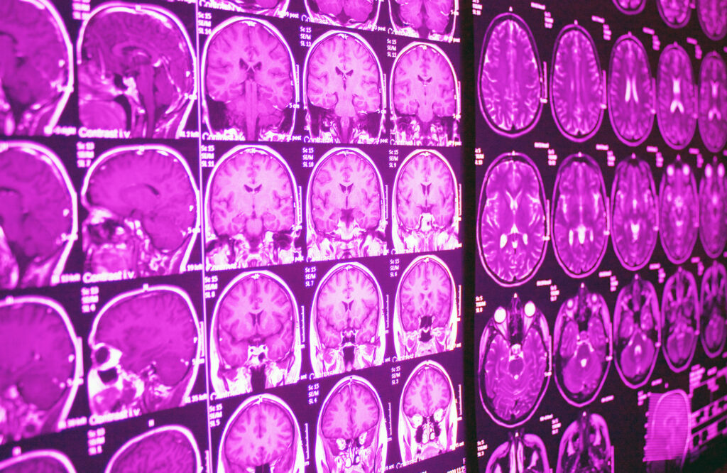
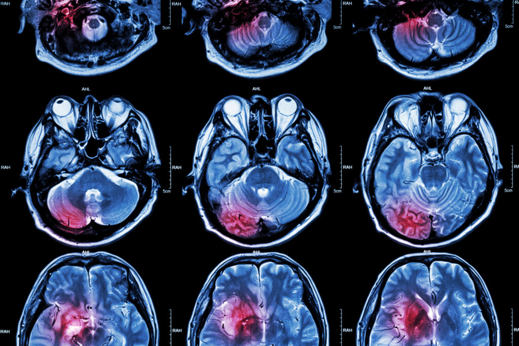
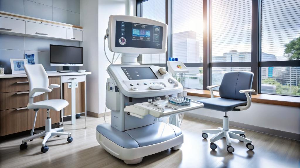
X-ray imaging has been a foundation of medical diagnostics for more than a century, yet the technology behind it continues to advance at a remarkable pace. Hospitals, clinics, and research centres now have access to systems that deliver sharper images, lower radiation doses, and faster workflows than ever before. As science and engineering progress, X-ray machines are moving far beyond their traditional form, gaining new capabilities that promise significant improvements for both clinicians and patients.
This article explores the latest developments that are redefining what X-ray machines can achieve, covering detector technology, image acquisition methods, digital integration, artificial intelligence, and the influence of industrial innovation on healthcare imaging.
Ultra-Low Dose Detectors and Safer Imaging
One of the strongest drivers in modern X-ray system development is patient safety. Reducing radiation exposure has long been a priority, especially in settings where individuals require repeated imaging. Recent scientific breakthroughs in detector design have unlocked new opportunities to achieve high-quality images at much lower doses.
A key advancement is the development of detectors with far greater sensitivity to incoming photons. These sensors capture more information from shorter exposures, enabling the machine to produce clear clinical images while placing less strain on the patient. Several research teams are investigating materials and detector architectures that enhance signal collection, reduce noise, and improve conversion efficiency.
A particularly promising direction involves “cascade-engineered” detectors, which use structured layers to amplify the signal. The result is better image quality with reduced exposure settings, offering safety benefits without compromising diagnostic confidence. As these technologies transition into commercial systems, clinicians will be able to perform routine radiographs with reduced risk for vulnerable groups, such as children, pregnant patients, and long-term monitoring cohorts.
Multi-Contrast and Phase-Contrast X-Ray Imaging
Traditional radiography relies on attenuation contrast—the degree to which tissues absorb or scatter X-rays. While this form of contrast is useful for detecting fractures, lung pathology, or large abnormalities, it has limitations when dealing with soft tissues that share similar attenuation profiles.
New imaging approaches are being developed to expand the information available from X-ray beams. Multi-contrast imaging captures multiple types of information simultaneously, including attenuation, phase shift, and dark-field signals. Phase-contrast imaging, for example, detects changes in the phase of X-rays as they pass through tissue. This is sensitive to structural differences at a scale far below that of conventional methods.
The potential applications are wide-ranging. In medical diagnostics, phase-contrast imaging can highlight subtle changes in soft tissues, offering improved visualisation of early-stage disease. In security and industrial testing—areas that often pioneer these methods—multi-contrast systems have demonstrated outstanding performance in classifying concealed or complex materials. As this technology becomes more accessible, medical radiography stands to benefit from clearer, more informative images that support earlier and more accurate diagnosis.
Advances in Digital Radiography
Digital radiography (DR) continues to evolve, gradually replacing analogue and film-based systems across healthcare settings. Modern DR units use flat-panel detectors with enhanced resolution and dynamic range, offering sharper images and faster capture times. The shift to fully digital systems streamlines the imaging workflow, reduces repeat scans, and improves data portability.
Current generations of DR detectors use direct conversion materials such as amorphous selenium or cadmium telluride, which convert X-ray photons directly into electrical signals. Direct conversion reduces blur and improves detail, especially at low doses. These detectors sit at the heart of many new X-ray machines, contributing to consistent, dependable imaging that supports confident clinical decisions.
Digital systems also facilitate seamless integration with hospital information systems, picture archiving and communication systems (PACS), and cloud-based storage solutions. This allows clinicians to retrieve images instantly, share them across departments, and work more efficiently during urgent cases.
The Rise of 3D and Volumetric X-Ray Capabilities
While computed tomography (CT) is the established method for three-dimensional imaging, new X-ray technologies are beginning to bridge the gap between traditional radiography and volumetric imaging. Researchers are exploring X-ray systems that capture information from multiple angles or energy levels, enabling reconstructions that offer depth cues and a richer understanding of internal structures.
One area gaining traction involves multi-energy imaging, where different X-ray energies interact uniquely with tissues. Combining these datasets can produce enhanced images that highlight features invisible to standard radiographs. Another direction involves advanced algorithms capable of generating quasi-3D information from limited projections. These capabilities could eventually allow clinicians to assess certain conditions more accurately without resorting to full CT scans.
Although still emerging, these techniques point to a future in which even basic X-ray units provide significantly more information than current systems.
Artificial Intelligence as a Core Component of Modern X-Ray Systems
The integration of artificial intelligence into X-ray imaging has progressed rapidly. AI algorithms assist radiographers and radiologists throughout the imaging chain—from positioning and exposure selection to image analysis and reporting.
On the acquisition side, AI can automatically identify the imaged body part and adjust exposure settings accordingly. This reduces variability between operators and lowers the likelihood of errors or repeat scans. During interpretation, AI can detect features such as lung nodules, fractures, cardiomegaly, pneumothorax, and other abnormalities at a speed impossible for manual review.
Importantly, AI is not replacing radiologists. Instead, it functions as a tool that supports clinicians by highlighting areas of interest, prioritising urgent cases, and reducing the cognitive load in busy clinical environments. The result is a more streamlined workflow, faster turnaround times, and potentially earlier diagnosis for time-critical conditions.
Cross-Domain Innovation: How Industrial Imaging Shapes Medical Progress
A great deal of innovation in X-ray technology originates outside of medicine. Security scanning, non-destructive testing, material science, and industrial inspection frequently adopt cutting-edge imaging methods long before they reach hospital settings. These domains provide a testbed for new detector materials, contrast modes, and computational techniques.
For example, imaging approaches used to identify concealed weapons or inspect aircraft components have contributed to advances in multi-contrast X-ray imaging. Industrial detectors designed to capture fine structural details with minimal noise are now influencing medical detector design. These cross-domain exchanges accelerate progress, bringing new ideas and breakthroughs into clinical environments more quickly than would otherwise be possible.
The Future of X-Ray Machines
The next wave of progress in X-ray technology is likely to focus on portability, personalisation, and improved data intelligence. Portable and even handheld X-ray units are becoming more capable, allowing imaging in emergency situations, remote areas, or care-at-home settings. Detectors will continue to improve in sensitivity and durability, further reducing dose.
AI will deepen its presence, providing instant pre-screening, automating administrative tasks, and supporting training for less-experienced staff. Meanwhile, advances in source technology may eventually lead to compact systems with higher coherence or tunable X-ray beams.
As these developments continue, X-ray imaging will remain central to diagnostics, offering safer, sharper, and more informative results for patients and clinicians alike.
Disclaimer
The information provided in this article is intended for general knowledge and educational purposes only. It is not a substitute for professional medical advice, diagnosis, or treatment. Readers should always seek guidance from qualified healthcare professionals regarding medical decisions or concerns. While every effort has been made to ensure accuracy at the time of publication, developments in medical imaging and related technologies may evolve, and Open MedScience does not guarantee the completeness or current validity of the content. Open MedScience is not responsible for any actions taken based on the information presented herein.


