Summary: Asthma affects hundreds of millions of people worldwide and remains a significant public health challenge. The primary treatment involves inhaled medicines delivered through aerosol devices such as pressurised metered dose inhalers, dry powder inhalers, soft mist inhalers, and nebulisers. The effectiveness of these devices depends on drug formulation, particle size, patient technique, and adherence. Medical imaging, although not a routine part of standard asthma care, plays a crucial role in research and advanced clinical settings. Techniques such as high-resolution CT, hyperpolarised gas MRI, and nuclear medicine ventilation studies provide detailed insights into airway obstruction, airflow patterns, and drug deposition. By linking aerosol delivery systems with imaging technologies, clinicians and researchers can optimise device performance, enhance patient outcomes, and move towards more personalised approaches to asthma management.
Keywords: Asthma, Aerosol devices, Inhalers, Medical imaging, Drug deposition, Personalised therapy.
Introduction
Asthma is one of the most common chronic respiratory conditions worldwide, affecting more than 300 million people. The disease is characterised by variable airway inflammation, bronchoconstriction, and reversible airflow obstruction. While treatment strategies include oral therapies and biologic agents for severe cases, the mainstay of management remains the use of inhaled medicines delivered directly to the lungs.
Delivering drugs in aerosol form allows high local concentrations in the airways while reducing systemic side effects. However, the effectiveness of inhaled therapy depends not only on the drug itself, but also on the device used and the patient’s technique. At the same time, medical imaging plays a crucial role in understanding both the disease process and the distribution of aerosol particles within the lungs. By combining inhaler technology with imaging insights, clinicians and researchers can refine asthma management and explore more personalised approaches to care.
Aerosol Devices for Asthma
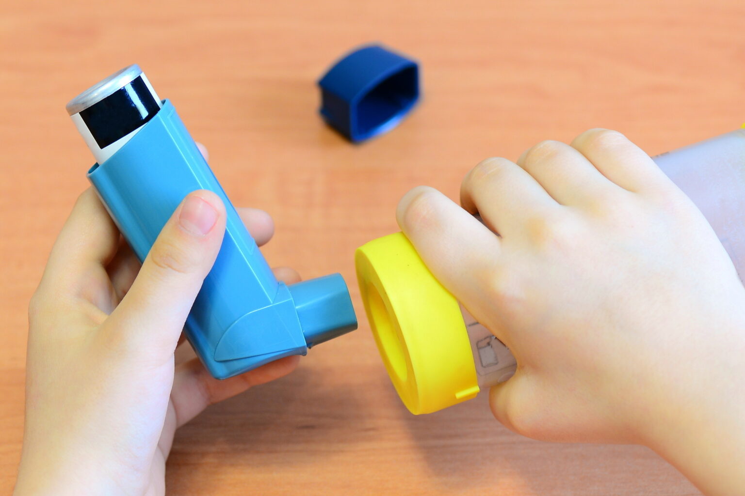
Pressurised Metered Dose Inhalers (pMDIs)
The pMDI is one of the most widely used inhaler types. It contains a pressurised canister that delivers a precise dose of medication in aerosol form when actuated. Patients inhale the released puff while coordinating their breathing with the device.
The advantages of pMDIs include portability, consistent dosing, and relatively low cost. However, poor technique — such as failing to shake the device, inhaling too late or too quickly, or not holding the breath afterwards — can significantly reduce drug delivery to the lungs. Spacers or holding chambers are often prescribed to improve deposition and make timing less critical.
Dry Powder Inhalers (DPIs)
DPIs contain medication as a fine powder, which is dispersed by the patient’s own inspiratory effort. Unlike pMDIs, they do not require coordination between actuation and inhalation. Instead, patients must generate sufficient inspiratory flow to aerosolise the powder effectively.
These devices are breath-actuated, which reduces timing errors, but they may be less suitable for very young children, the elderly, or those with severe airflow obstruction who cannot inhale forcefully.
Soft Mist Inhalers (SMIs)
SMIs are a more recent development, producing a slow-moving aerosol cloud that lasts longer than the brief puff from a pMDI. The softer mist increases the likelihood of drug particles penetrating deep into the lungs, while also reducing deposition in the oropharynx. Patients generally find them easier to use, and clinical studies suggest improved lung deposition compared with traditional inhalers.
Nebulisers
Nebulisers convert liquid medication into a fine mist over several minutes, inhaled via a mouthpiece or facemask. They are particularly useful for patients who are unable to use handheld inhalers effectively, such as young children, individuals experiencing severe exacerbations, or those in hospital settings.
While effective, nebulisers are less convenient for routine daily use because they require power, are bulkier, and have longer administration times.
Clinical Considerations
The success of aerosol therapy depends on several factors:
- Particle size: Optimal deposition occurs when particles are between 1–5 micrometres. Larger particles are trapped in the upper airways; smaller particles may be exhaled without depositing.
- Inhaler technique: Incorrect use remains one of the greatest barriers to effective therapy. Education and repeated demonstration are essential.
- Adherence: Many patients underuse or overuse inhalers. Devices with dose counters or digital monitoring features are being introduced to address this issue.
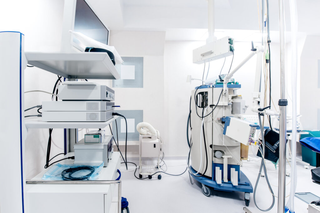
Medical Imaging in Asthma
Although asthma is primarily a clinical diagnosis, imaging plays a significant role in research, the management of severe cases, and the exclusion of alternative conditions.
Chest X-ray
Often performed at initial presentation to rule out infection, pneumothorax, or other structural abnormalities. For typical asthma, chest X-rays are usually normal.
Computed Tomography (CT)
High-resolution CT (HRCT) provides detailed images of the lungs and airways. In patients with chronic or severe asthma, CT scans may reveal bronchial wall thickening, air trapping, or mucus plugging. Quantitative CT can measure airway dimensions, helping to assess remodelling over time.
Magnetic Resonance Imaging (MRI)
Conventional MRI is limited in lung imaging due to the low proton density of air spaces. However, newer techniques using inhaled hyperpolarised gases such as helium-3 and xenon-129 provide striking images of ventilation distribution. These methods can map regional airflow obstruction in exquisite detail, without ionising radiation.
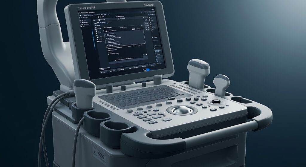
Nuclear Medicine Ventilation Imaging
Radionuclide techniques involve inhaling radiolabelled aerosols or gases to trace airflow through the lungs. Planar scintigraphy, SPECT (Single Photon Emission Computed Tomography), and PET (Positron Emission Tomography) have all been used to study regional ventilation defects. These tools offer a functional view of lung performance, complementing the structural details provided by CT or MRI.
Functional and Hybrid Imaging
Modern approaches often combine structural and functional imaging, offering a comprehensive view of how airways behave in health and disease. This integration is particularly valuable in asthma research, where airway obstruction is often patchy and dynamic.
Linking Aerosol Devices and Imaging
Medical imaging has become crucial in understanding how inhaled therapies function within the lungs. By radiolabelling inhaled drugs or simulating aerosol particles, researchers can directly visualise deposition patterns.
- Scintigraphy and SPECT studies: These have been widely used to show how different inhaler types, spacers, and inhalation techniques affect drug distribution. They reveal that poor coordination with pMDIs leads to much higher oropharyngeal deposition, whereas spacers increase lung delivery.
- PET imaging: When drugs or particles are labelled with positron-emitting isotopes, PET can provide high-resolution, three-dimensional maps of deposition and clearance over time.
- Hyperpolarised gas MRI: By mapping areas of reduced ventilation, researchers can see how disease heterogeneity affects inhaler performance. For example, obstructed airways may prevent aerosol particles from reaching certain lung regions.
- Device optimisation: Imaging data helps engineers and clinicians refine inhaler designs, particle size distributions, and delivery strategies, ensuring that more of the drug reaches the intended target.
Together, these methods demonstrate that imaging is not just a diagnostic tool but a key partner in therapeutic development.
Future Directions
The intersection of inhaler technology and imaging is opening exciting possibilities:
- Personalised therapy: Imaging may guide which inhaler type is most effective for a particular patient, depending on their airway anatomy and disease pattern.
- Digital health integration: Smart inhalers that monitor usage could one day be combined with imaging data to track how adherence relates to airway changes.
- AI and computational modelling: Advanced algorithms can predict particle behaviour within individual lungs, validated by imaging studies. This could allow inhaler therapy to be tailored as precisely as chemotherapy dosing in oncology.
- Reduced radiation strategies: As MRI and other non-ionising methods advance, the ability to study aerosol deposition safely in children and young adults will grow.
Conclusion
Aerosol devices remain the cornerstone of asthma management, providing direct delivery of medications to the airways where they are most needed. However, their effectiveness depends on the interplay between drug formulation, device mechanics, patient technique, and underlying disease.
Medical imaging, although not used routinely in everyday asthma care, provides unique insights into lung structure and function. From visualising airflow obstruction with hyperpolarised gases to mapping drug deposition with nuclear medicine, imaging enables clinicians and researchers to understand how therapies actually perform inside the lungs.
As technology advances, the synergy between inhaler devices and imaging promises more precise, personalised asthma treatment. What began as a simple puff of aerosol is evolving into a carefully engineered partnership between pharmaceutical science, medical imaging, and patient care.
Disclaimer
The information presented in this article, “Aerosol Devices for Asthma and Aspects of Medical Imaging”, is intended solely for general educational and informational purposes. It does not constitute medical advice, diagnosis, or treatment, and should not be relied upon as a substitute for consultation with a qualified healthcare professional. Readers should always seek the guidance of a doctor, pharmacist, or respiratory specialist regarding any medical condition, treatment options, or the use of inhaler or imaging technologies.
Open MedScience and the article’s authors make no representations or warranties regarding the accuracy, completeness, or applicability of the information contained herein. While every effort has been made to ensure that the content is based on current scientific understanding at the time of publication, medical knowledge and technologies evolve continuously. Therefore, the authors and Open MedScience accept no liability for any loss, injury, or damage arising from the use of or reliance on the material provided.
References to specific medical devices, imaging methods, or technologies are for illustrative purposes only and do not imply endorsement or commercial association.
home » blog » medical devices »