Positron Emission Tomography (PET scanners) are medical imaging machines that use radiotracers to produce 3-D images of the human body’s internal structures and functions. These radiotracers can be injected into the patient’s bloodstream, inhaled, or swallowed. As the radiotracer travels through the body, it emits positively charged particles (positrons), which collide with negatively charged electrons in the body’s tissues. This collision produces gamma rays, detected by the PET scanner and used to create detailed anatomical images.
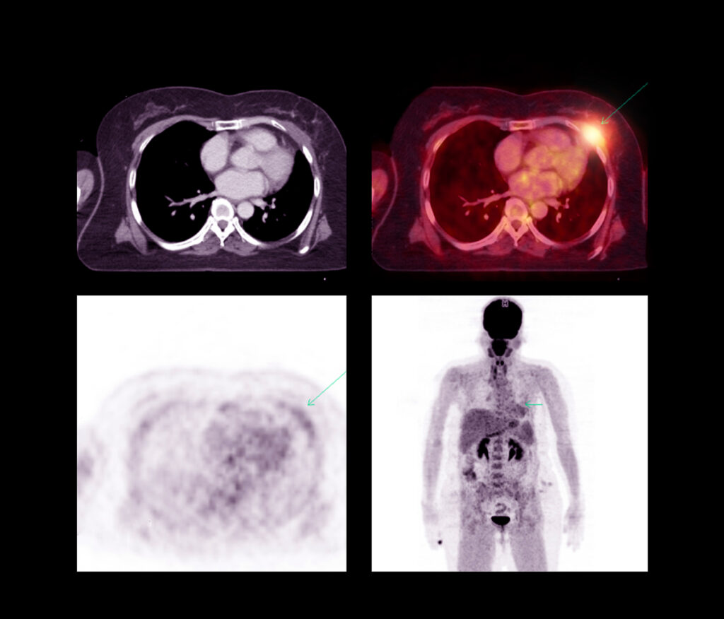
PET scanners diagnose and monitor various medical conditions, including cancer, heart disease, and brain disorders. They can also assess treatments’ effectiveness and guide surgical procedures. In addition, PET scans are often combined with CT (computed tomography) or MRI (magnetic resonance imaging) scans to provide even more detailed information about the body’s structures and functions.
The most used radioisotope in PET scanning is fluorine-18 (t1/2= 109.77 mins), which creates a tracer molecule called fluorodeoxyglucose (2-deoxy-2-[18F]fluoro-D-glucose) known as FDG.
FDG is a form of glucose modified to include a radioisotope of fluorine. When injected into the body, FDG is taken up by cells that use glucose as an energy source, such as cancer cells. As the FDG accumulates in these cells, it emits positrons, which collide with electrons in the surrounding tissue to produce gamma rays detected by the PET scanner.
Other radioisotopes used in PET scanning include carbon-11 (t1/2= 20.34 mins), nitrogen-13 (t1/2= 9.965 mins), and oxygen-15 (t1/2= 2.03 mins). These radioisotopes are used to create tracers that target specific biochemical processes in the body, such as blood flow or oxygen consumption.
It’s important to note that the radioisotopes used in PET scanning have short half-lives, meaning they decay rapidly and lose their radioactivity within a short period of time. This minimises the risk of long-term radiation exposure to the patient.
Positron Emission Tomography: Principles and Applications
Several advanced PET scanners are currently available, each with unique features and capabilities, for example:
- Digital photon counting (DPC) PET scanners use advanced detectors to detect individual photons emitted by the radioactive tracer. This allows for higher spatial resolution, faster acquisition times, and improved sensitivity compared to traditional PET scanners.
- Time-of-flight (TOF) PET scanners use advanced detectors to measure the time gamma rays travel from the tracer to the detector. By measuring the time of flight, TOF PET scanners can improve the accuracy and quality of the images produced.
- Total-body PET scanners can image the entire body in a single scan, allowing for more comprehensive disease and treatment response assessments.
- Hybrid PET/MRI scanners combine PET and MRI technologies, allowing for the simultaneous acquisition of anatomical and functional information. This provides a complete picture of the anatomical structures and functions.
- Preclinical PET scanners are designed for animal research and drug development. They offer higher spatial resolution and greater sensitivity than clinical PET scanners, allowing researchers to study disease processes at the cellular and molecular levels.
The field of PET scanning is rapidly evolving, and new technologies and advancements are constantly being developed.
Neuroimaging with PET: Advances in Brain Disorders
PET imaging is an important tool in neurology for assessing brain structure and function. This diagnostic imaging tool can detect the accumulation of amyloid plaques and tau tangles in the brain, characteristic of Alzheimer’s disease. This can help with early diagnosis and monitoring of disease progression. It can also measure dopamine levels in the brain, which are depleted in Parkinson’s disease. This can help with the diagnosis and monitoring of disease progression. Furthermore, a PET examination can identify the brain’s specific regions responsible for seizures, which can help with surgical planning and treatment decisions. In addition, PET imaging can be used to assess blood flow and metabolic activity in the brain following a stroke.
Hybrid Imaging: Integrating PET with MRI and CT
PET scanner hybrids are machines that combine PET technology with other imaging modalities, such as CT, MRI, or SPECT (single photon emission computed tomography). These hybrid scanners offer several advantages over conventional PET scanners, including:
- Improved anatomical localisation by combining PET with other imaging modalities; hybrid scanners can provide more precise anatomical localisation of the areas of interest.
- Hybrid scanners can provide complementary information from multiple imaging modalities, improving the accuracy of diagnoses and treatment planning.
- By combining PET with other imaging modalities, hybrid scanners can reduce the total radiation exposure to the patient compared to separate PET and CT scans.
- Hybrid scanners can perform multiple imaging modalities in a single session, reducing the time and cost of separate imaging sessions.
PET scanner hybrids include PET/CT, PET/MRI, and SPECT/CT. PET/CT combines PET and CT technologies to provide functional and anatomical information, making it particularly useful for cancer imaging. PET/MRI combines PET and MRI technologies to provide detailed anatomical and functional information with high soft tissue contrast, making it particularly useful for brain and musculoskeletal imaging. Finally, SPECT/CT combines SPECT and CT technologies to provide functional and anatomical information, making it particularly useful for cardiac imaging and bone scans.
PET scanner hybrids are powerful tools in medical imaging that allow for more precise and accurate diagnoses while reducing radiation exposure and increasing efficiency.
Milestones in PET Technology: Innovations That Shaped Modern Imaging
The development of PET imaging was a collaborative effort among several pioneers in physics, chemistry, and medicine. Some of the key figures involved in the development of PET imaging include:
- Gordon Brownell, in the 1950s, began researching the use of positron-emitting isotopes for medical imaging.
- Michel Ter-Pogossian collaborated with Brownell to develop the first PET scanner in the 1970s.
- Edward Hoffman helped to refine PET technology and develop new radiopharmaceuticals for use in PET imaging.
- David Kuhl developed new image reconstruction and interpretation techniques in PET imaging.
- Sami Shihab pioneered PET imaging in oncology, particularly in diagnosing and treating lung cancer.
These pioneers and other researchers develop and refined PET imaging technology, paving the way for its widespread use in modern medicine.
Next-Generation PET Scanners: A Glimpse into the Future of Molecular Imaging
Siemens Biograph PET/CT is a popular model of PET scanner manufactured by Siemens Healthineers, a global medical technology company headquartered in Germany. The Biograph PET/CT combines two imaging technologies, PET and CT (Computed tomography), into one device, allowing for more comprehensive body imaging.
This hybrid scanner is designed to provide high-resolution images with excellent contrast, helping to accurately detect and diagnose various medical conditions, including cancer, neurological disorders, and cardiovascular disease. In addition, the scanner uses a low radiation dose to produce images, making it safe for patients.
The Siemens Biograph PET/CT also has advanced software for efficient image reconstruction, analysis, and image sharing with other medical professionals. This makes it a valuable tool for medical research and clinical studies.
The GE Discovery PET/CT can produce detailed images of the body’s function and structure and help to diagnose cancer, heart disease, and neurological disorders.
The Philips Ingenuity TF PET/CT allows healthcare professionals to diagnose and treat various medical conditions, including cancer, heart disease, and neurological disorders. It has several advanced features, including time-of-flight PET technology, which helps improve image quality and reduce scan times, and a large bore, allowing larger patient imaging.
The Toshiba Celesteion PET/CT has several advanced features, including a high-speed CT scanner and a large detector ring for PET imaging, which can help reduce scan times and improve image quality.
The Mediso AnyScan PET/CT is a medical imaging scanner that can produce highly detailed images that provide information about the body’s function and structure. It has advanced features, including a high-resolution PET detector, a low-dose CT scanner, and a patient-friendly design that can help improve patient comfort during the scanning process.
Hitachi PET/CT scanners allow for imaging of larger patients, and a 64-slice CT scanner can help reduce scan times and improve image quality.
The NeuroLogica Quantum PET/CT has several advanced features, including a compact design, which allows for easier installation in smaller spaces, and a fast PET detector, which can help reduce scan times and improve image quality.
The Cubresa NuPET™ scanner is a preclinical medical imaging device that can produce highly detailed images that provide information about the body’s function and structure. This allows researchers to understand the underlying mechanisms of diseases better and develop new therapies to treat them.
Also, the Cubresa NuPET™ scanner has several advanced features, including a compact design, which allows for easier installation in smaller spaces, and a high-sensitivity PET detector, which can help reduce the amount of radioactive material needed for imaging. The device is used primarily in preclinical research and drug development and is considered a valuable tool in medical research.
The Raycan α-PET scanner uses silicon photomultiplier (SiPM) detectors, which are highly sensitive to light and can detect even very small amounts of radiation. This allows for a higher level of accuracy and sensitivity in PET imaging. The scanner also has a compact design, which allows for easier installation in smaller spaces and a fast acquisition time, which can help reduce scan times and improve patient comfort. The Raycan α-PET scanner is used primarily in research and clinical settings, particularly in diagnosing and treating cancer, neurological disorders, and cardiovascular disease.
The United Imaging uMI 550 PET/CT scanner has several advanced features, including a large bore, which allows for imaging of larger patients, and a high-definition digital detector, which can help reduce scan times and improve image quality. The device is used in hospitals and imaging centres worldwide and is considered a valuable tool in medical imaging. Additionally, the uMI 550 PET/CT has several advanced software tools for image reconstruction, quantitative analysis, and artificial intelligence-based interpretation, making it a powerful tool for clinical and research applications.
Total-Body PET/CT Imaging: Enhancing Diagnosis and Treatment Planning
The PET/CT Explorer scanner is a groundbreaking total-body imaging system that combines the functional information of PET with the anatomical details provided by CT. With its extended field of view, the PET/CT Explorer delivers enhanced sensitivity, enabling faster scans, reduced radiation doses, and improved patient diagnostic accuracy.
This innovative technology can transform clinical practice and research by providing a more comprehensive understanding of diseases and their progression. It is particularly beneficial for detecting cancer, monitoring treatment response, and assessing cardiovascular and neurological conditions.
Furthermore, the PET/CT Explorer scanner can open new opportunities for research in pharmacokinetics, immunology, and stem cell therapy. This advanced imaging system paves the way for more personalised medicine and improved patient outcomes by providing a holistic view of the human body.
Disclaimer
The information provided in this publication is intended for educational and informational purposes only. It is not a substitute for professional medical advice, diagnosis, or treatment. Readers should consult qualified healthcare professionals with any questions or concerns regarding their health or any medical condition.
Although every effort has been made to ensure the accuracy and reliability of the information presented, no guarantee is given regarding its completeness, timeliness, or applicability to individual cases. The technologies, radiotracers, and diagnostic methods described herein are subject to ongoing research, clinical evaluation, and regulatory approval, and their availability may vary depending on location and healthcare infrastructure.
Mention of specific commercial products, manufacturers, or scanner models does not imply endorsement. All product names and trademarks are the property of their respective owners.
Use of any information from this publication is at the reader’s own risk. Neither the authors nor the publishers accept any responsibility for any consequences arising from the use or misuse of the content.
home »