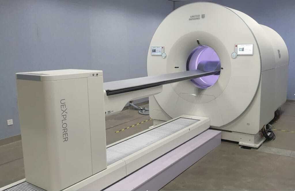
EXPLORER, the world’s first medical imaging scanner to produce a 3-D picture of the whole human body.
EXPLORER Total Body PET scanner
The EXPLORER Whole Body Scanner and its capabilities reminded me about the ‘Fantastic Voyage’. The Fantastic Voyage was a 1966 American science fiction film about a submarine crew who were shrunk to a microscopic size. Subsequently, they ventured into the human body of an injured scientist to repair damage to his brain. The synergy is the ability of the EXPLORER to track a radiopharmaceutical through the human body and locate the disease state in real-time.
However, future PET/CT hybrid scanners must be able to use most of the ‘PET signal’. At present, all PET scanners are limited by low numbers of detected photons and high dosages of radiation. Nevertheless, some of these challenges have been addressed by the EXPLORER.
The EXPLORER, the world’s first medical imaging scanner, can produce a 3-D picture of the whole human body in one session and, since its development 13 years ago, has recently produced its first scans. This next-generation hybrid machine was created by project leaders Simon Cherry and Ramsey Badawi in partnership with United Imaging Healthcare (UIH) and other organisations at a cost of over $15 m. The EXPLORER combines the imaging modalities of PET Imaging (PET) with X-ray computed tomography (CT).
This revolutionary scanner can generate a whole body image within one second and is equipped with advanced video technology to track radiopharmaceuticals as they have a Fantastic Voyage through the entire body!
EXPLORER PET/CT scanner design and features
Video Radiotracing
Video technology can show the distribution of the positron emission radiotracer FDG (2-deoxy-2-[18F]fluoro-D-glucose) upon in vivo administration into the patient’s vein, which is captured in real time by the EXPLORER scanner. These detailed 3-D images produced from the dynamic sequence are not shown on standard PET scanners.
- The PET radiotracer is injected into the patient’s vein, and after a short time, it travels to the heart. At this point, the radiotracer is dispersed throughout the arteries to all organs of the body.
- At approximately 3 minutes, a percentage of the radiotracer is excreted from the kidneys into the bladder.
- Accumulation of the FDG can be detected in the brain and heart, including the liver, over this interval.
- The EXPLORER scanner is capable of investigating the metabolism and excretion of a radiolabelled version of the drug throughout the entire human body.
- There is a fine balance between image quality, acquisition time, and injected radiation dose. This combination will vary between different applications, but in most instances, the EXPLORER can scan faster and obtain images at a much lower radiation dose.
In essence, the EXPLORER project team has accomplished the ability of a medical imaging scanner to capture a 3-D image of the whole human body, which has never been achieved before. This achievement will drive applications towards improving diagnostic medical imaging and developing specific radiopharmaceuticals to track the progression of the disease state.
Future PET/CT scanners will benefit a patient’s treatment plans because these EXPLORER machines will provide higher-quality diagnostic PET scans, which are not currently available. The EXPLORER is able to scan up to 40 times faster than current PET scanners and deliver a diagnostic scan of the whole body within 30 seconds.
The primary benefit of the EXPLORER is that it can scan a patient using a radiation dose that is 40 times less than what is used in conventional PET scanners. This significant reduction in the dose will allow for repeat scans especially for children where the cumulative radiation dose is an important factor to be considered.
For the first time, the EXPLORER can evaluate what is happening in all organs and tissues of the body. For example, it has the scope to quantitatively measure blood flow and investigate the mechanism of how the body takes up glucose in the human body. The EXPLORER will be the apex imaging modality in the study of cancer, causes of inflammation and infection as well as immunological and metabolic disorders of the disease state.
The substantial increase in the EXPLORER sensitivity will enable it to:
- produce more reliable images
- obtain images by scanning at very low radiation dosages
- rapidly scan within 1 minute
- evaluate the PET radiopharmaceutical after a more significant time interval from an injection of the radiotracer
The EXPLORER total-body imaging applications include:
- fast scanning, which requires no anaesthesia
- detection of infections and chronic diseases such as cancer
- investigation of cell-based therapies
- evaluation of drug pharmacokinetics in all organs of the body
- study of metabolic disorders and autoimmune diseases
- toxicological research
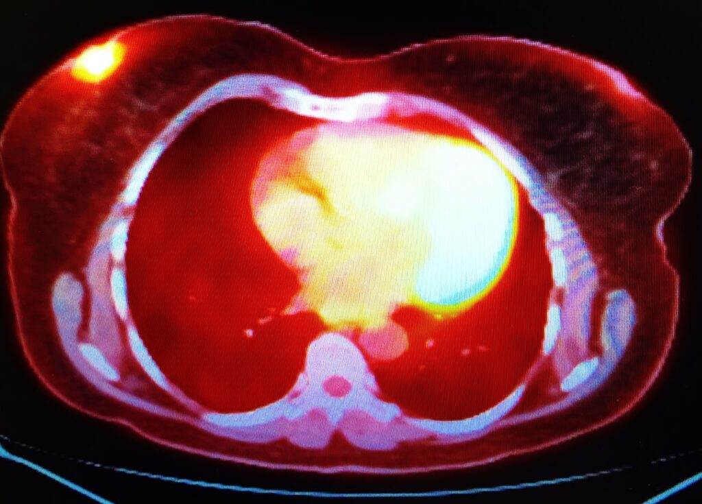
- Generation of coronal, sagittal and axial slices of the human body.
- research into the endocrine and immunological signalling implicated in a range of disorders, including irritable bowel syndrome
- Identify the pharmacokinetics (pk) of new medicines in all organs of the body at lower mass and radiation dosages of 100 µSv.
- used in drug development and toxicology studies
EXPLORER is a scanner that is capable of generating 3-D images of the whole human body.
The data produced by the whole body scanner will be used to extract detailed information regarding metabolism, radiopharmaceuticals and drug-receptor interactions.
The EXPLORER PET/CT scanner specifications
The future
The anatometabolic imaging modalities combined SPECT/CT, PET/CT, and PET/MR imaging are available for clinical use. These hybrid imaging scanners can provide advanced information on the disease state by using big data and computer-based models for disease and therapy, including response prediction. The EXPLORER has set the benchmark for the next generation of PET/CT scanners.
Future developments will have to address clinical applications regarding ultra-fast paediatric scans and limit anaesthesia use. The EXPLORER also allows for low-dose follow-up scanning, especially in paediatric oncology. The next generation of whole-body scanners must be able to produce rapid higher resolution images using low dose radiation and machines to generate total-body dynamic images with high temporal resolution.
Disclaimer
The content of this article is intended for informational and educational purposes only. It does not constitute medical advice, diagnosis, or treatment, and should not be relied upon as such. The information presented reflects the views of the author(s) at the time of publication and may not reflect the most current research or clinical developments. Readers should consult with qualified healthcare professionals for specific medical guidance or concerns.
While every effort has been made to ensure the accuracy of the details provided, Open Medscience does not guarantee the completeness or reliability of the information, particularly in relation to clinical applications, safety, or performance outcomes of the EXPLORER PET/CT scanner. Open Medscience and its contributors accept no responsibility for any loss, damage, or harm arising from the use or interpretation of the content herein.
Any reference to third-party organisations, researchers, or technologies is made for context and does not imply endorsement by Open Medscience.
home » blog » nuclear medicine imaging »